| 영문 | retinal detachment | 한글 | 망막 박리 |
|---|---|---|---|
| 설명 | 카메라에 있어서 필름에 해당하는 눈의 망막은 크게 두 개의 층으로 나눌 수가 있다. 안쪽에 있는 실제의 빛을 감지하는 감각층과 바깥쪽의 외부의 빛을 차단하는 색소상피층이 그것인데 그 사이에는 잠재적인 공간이 있어서 떨어지기가 쉽다. 이 사이가 떨어지면 망막의 감각층이 망막의 색소상피층과 분리되는데 이것을 망막박리라고 한다. 이 망막의 박리에는 여러 가지 원인이 있지만 감각층의 망막에 작은 구멍인 열공(break)에 의해서 그곳으로 눈속을 채우고 있는 액체가 흘러 들어가서 생기는 망막의 박리를 열공성 망막박리(rhegmatogenous retinal detachment)라 하고, 안구의 병터에 의해서 안구내에 섬유조직이 생기고 그것이 망막의 감각층을 잡아 끌어서 망막이 박리되는 견인성 망막박리(traction retinal detachment) 및 망막의 2개의 층에 삼출액이 괴어서 생기는 삼출성 망막박리(exudative retinal detachment) 등 열공에 의해서 생기는 망막박리가 아닌 것을 비열공성 망막박리(nonrhegmatogenous retinal detachment)라고 말한다. |
||
| 영문 | muscle cell(=muscle fiber) | 한글 | 근육세포 |
|---|---|---|---|
| 설명 | 근육은 수의근(의식에 의해서 조절이 가능한 근육: 예를 들어 다리, 팔, 얼굴근육 등)과 불수의근(의식과 무관하게 조절하지 않아도 움직이는 근육: 예를 들어 심장근, 소화기관에 분포하는 근육 등)으로 나누어지며, 근육세포의 모양은 다음과 같다(수의근, 불수의근 외에 심장에 있는 심장근은 불수의근에 해당하지만 그 모양은 수의근과 같아 따로 분류한다).  |
||
| 영문 | white blood cell(WBC), leukocyte | 한글 | 백혈구 |
|---|---|---|---|
| 설명 | 혈액내에 골수구계세포와 림프계세포, 단핵구계세포를 모두 통틀어 말한다. 백혈구의 증가가 있으면 대개 감염이 있거나, 혹은 탈수현상이 있음을 의미한다. 또한 지나친 백혈구수의 감소는 인체내 면역기능이 떨어져 있음을 의미하며, 다른 질병에 의해 나타나는 이차적인 현상이 아닌지 꼭 진단을 받아보아야 한다. |
||
| 영문 | mast cell | 한글 | 비만 세포 |
|---|---|---|---|
| 설명 | 동물의 결합 조직 가운데 널리 분포하는 세포. 결합조직과 점막조직 내에 있는 호염기성 색소로 이염색성(metachromasia)을 나타내는 과립을 가진 방추형의 세포에 작은 둥근 핵을 가진다. 비만세포의 표면에는 IgE에 대한 수용체가 존재하며, 수용체에 결합한 IgE 분자들끼리 다가의 항원에 의해 서로 연결되면 비만세포 과립탈출 반응이 일어나, 히스타민, 세로토닌, 헤파린 등의 화학전달 물질이 방출되어, 즉시형 알레르기 반응 등의 증상을 일으킨다. 피부, 장막, 혈관 주위, 점막 주변에 있다. |
||
| 영문 | cell | 한글 | 세포 |
|---|---|---|---|
| 설명 | 생명체를 이루는 구조적, 기능적 단위. 핵의 유무에 따라서 유핵세포와 무핵세포로 나눌 수가 있다. 무핵세포란 핵이 없는 세포를 말한다. 핵이란 유전정보를 가지고 있는 염색체를 보관하는 곳인데 무핵세포에서는 염색체가 세포속에 그냥 노출되어 있다. 대개 세포 하나하나가 독립된 생명체의 역할을 하는 단세포생물로서 외부환경으로부터 자신을 보호할 수 있는 견고한 세포벽을 가지고 있다. 유핵세포란 핵을 가지고 있어서 유전정보가 있는 염색체가 세포의 다른 부분과 구분되어 핵속에 들어있다. 세포속에서 핵외의 부분(이를 세포질이라 한다)에는 여러 가지 세포의 소기관이 있어서 세포의 다양한 기능을 분담한다. ◈유핵세포의 기본 구조 1.세포막-세포를 둘러싸서 주위환경과 분리하여 세포의 내환경을 유지한다. 2.형질내세망(endoplasmic reticulum)-단위막으로 둘러쌓여 있는 불규칙한 망상구조이다. 이 망상구조의 내부를 소강, 소조라고 부른다. 여기에는 표면이 매끈한 무과립형질내세망과 표면이 불규칙한 과립형질내세망의 두 가지가 있다. 과립형질내세망의 경우는 표면에 단백질을 합성하는 리보솜이라는 것이 부착되어 있다. 이곳은 주로 세포밖으로 분비할 단백질을 합성하는 장소이다. 무과립형질내세망은 표면에 리보솜이 부착되어 있지 않은 것을 말하며, 이곳에서는 주로 해독작용, 글리코겐의 합성, 스테로이드호르몬의 합성 등이 일어난다. 3.리보솜-단백질을 합성하는 역할을 하는 곳이다.이것은 세포질에 존재하는 자유리보솜과 과립형질내세망에 부착이 되어 존재하는 부착리보솜의 두 가지로 나눈다. 자유리보솜은 주로 세포내에서 필요한 단백질을 만드는 역할을 하고 부착리보솜은 세포밖으로 분비할 단백질을 만드는 역할을 한다. 4.골지장치(Golgi apparatus)-핵주위에 분포하는 납작해진 주머니모양의 것이 중첩되어 형성된 층판 모양의 구조물로 과립형질내세망에서 생성되어 외부로 분비될 단백질을 모아서 농축, 포장하여 과립을 만드는 역할을 한다. 5.사립체(mitochondria)-구형, 난형의 긴 막대기 모양으로 크기가 다양한 구조물. 생물체의 에너지 저장물질인 ATP를 생산하는 역할을 한다. 또 세포와 다른 자신만의 유전정보를 가진 DNA, RNA를 가지고 있다. 모양, 크기가 세균과 비슷하며 자체증식성 등 독립된 생명체로서 필요한 요건을 갖추고 있어서 세포와 공생관계를 가진 독립된 세포로 생각하고 있다. 6.용해소체(lysosome)-작은 구형의 소체로 여러 가지 분해효소를 가진다. 세포외계에서 들어온 물질과 결합하여 그 물질들을 용해하는 역할을 하고, 오래된 세포소기관들을 제거하는 역할도 한다. 7.세포핵(nucleus)-구형, 난형으로 세포의 중심에 위치한다. 핵내에는 유전정보가 있는 물질인 염색체가 존재한다. 8.중심소체(centrosome)-핵주위에 존재하면서 핵의 분열시에 양쪽의 염색체를 당기는 작은 섬유를 만드는 역할을 하는 곳. 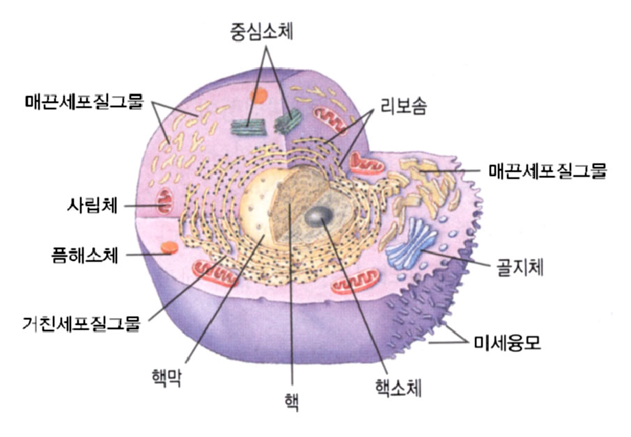 |
||
| RD | radial deviation; radiology department; rate difference; Raynaud disease; reaction of degeneration; ... |
|---|---|
| MC | mass casualties; mast cell; Master of Surgery [Lat. Magister Chirurgiae]; maximum concentration; Med... |
| RGC | radio-gas chromatography; remnant gastric cancer; retinal ganglion cell; right giant cell |
| ACC | accommodation; acetyl coenzyme A carboxylase; acinic cell carcinoma; acute care center; adenoid cyst... |
| ARC | accelerating rate calorimetry; acquired immunodeficiency syndrome-related complex; active renin conc... |
| BREC | bovine retinal endothelial cell |
|---|---|
| RGC | Retinal ganglion cell |
| ARN | Acute retinal necrosis |
| ARN | Acute retinal necrosis syndrome |
| ARC | Anomalous retinal correspondence |
| retinal ganglion cell | <pathology> A type of interneuron that conveys information from the retinal bipolar, horizontal and amacrine cells to the brain. (18 Nov 1997) |
|---|---|
| retinal pigmented epithelial cell | See: pigmented retinal epithelium, retina. (18 Nov 1997) |
| all-trans-retinal | The orange retinaldehyde resulting from the action of light on the rhodopsin of the retina, which converts the 11-cis-retinal component of the rhodopsin to all-trans-retinal plus opsin. Synonym: trans-retinal, visual yellow. (05 Mar 2000) |
| blood-retinal barrier | Specialised nonfenestrated tightly-joined endothelial cells that form a transport barrier for certain substances between the retinal capillaries and the retinal tissue. (12 Dec 1998) |
| central retinal artery occlusion | <ophthalmology> The sudden blockage of the retinal artery with a blood clot that commonly leads to a painless but irreversible blindness in that eye. (12 Jan 1998) |
| central retinal fovea | A depression in the centre of the macula retinae containing only cones and lacking blood vessels. Synonym: fovea centralis retinae, central pit. (05 Mar 2000) |
| central retinal vein occlusion | <ophthalmology> The sudden blockage of the retinal vein with blood clot that commonly leads to a painless irreversible blindness in that eye. (12 Jan 1998) |
| retinal | 1. <anatomy> Pertaining to the retina. 2. <biochemistry> The aldehyde of retinol, derived by the oxidative enzymatic splitting of absorbed dietary carotene and having vitamin A activity. In the retina, retinal combines with opsins to form visual pigments. One isomer, 11 cis retinal combines with opsin in the rods (scotopsin) to form rhodopsin or visual purple. Another, all trans retinal (trans r.), visual yellow, xanthopsin) results from the bleaching of rhodopsin by light, in which the 11 cis form is converted to the all trans form. Retinal also combines with opsins in the cones (photopsins) to form the three pigments responsible for colour vision. (18 Nov 1997) |
| retinal adaptation | Adjustment to degree of illumination. (05 Mar 2000) |
| retinal anlage tumour | A benign neoplasm of neuroectodermal origin that most often involves the anterior maxilla of infants in the first year of life. It presents clinically as a rapidly growing blue-black lesion producing a destructive radiolucency; histologically, it is characterised by small round undifferentiated tumour cells interspersed with larger polyhedral melanin-producing cells arranged in an alveolar configuration. Synonym: melanoameloblastoma, pigmented ameloblastoma, pigmented epulis, progonoma of jaw, retinal anlage tumour. (05 Mar 2000) |
| retinal artery | <anatomy, artery> Central retinal artery and its branches. It arises from the ophthalmic artery, pierces the optic nerve and runs through its centre, enters the eye through the porus opticus and branches to supply the retina. (12 Dec 1998) |
| retinal artery occlusion | Occlusion or closure of the central retinal artery causing sudden, usually nearly complete, loss of vision in one eye. Occlusion of the branch retinal artery causes sudden visual loss in only a portion of the visual field. (12 Dec 1998) |
| retinal blood vessels | The blood vasculature of the retina, including the branches and tributaries of the central retinal artery and vein, respectively, and the vascular circle of the optic nerve. Synonym: vasa sanguinea retinae. (05 Mar 2000) |
| retinal camera | An instrument for photographing the ocular fundus. (05 Mar 2000) |
| retinal cone | <ophthalmology, physiology> One of the two photoreceptor cell types in the vertebrate retina. In cones the photopigment is in invaginations of the cell membrane of the outer segment. Cones are less sensitive to light than rods, and are differentially sensitive to particular wavelengths of light and therefore important for colour vision. They provide vision with higher spatial and temporal acuity, and it is the combination of signals from cones with different pigments that facilitates colour vision. There are three types of cones, each type sensitive to red, green or blue. Present in large numbers in the fovea. (03 Jul 1999) |
제품명 |
판매사 |
보험코드 | 성분/함량 | 구분/보험급여 |
|---|
제품명 |
판매사 |
보험코드 | 성분/함량 | 구분/보험급여 |
|---|