
| 영문 | muscle cell(=muscle fiber) | 한글 | 근육세포 |
|---|---|---|---|
| 설명 | 근육은 수의근(의식에 의해서 조절이 가능한 근육: 예를 들어 다리, 팔, 얼굴근육 등)과 불수의근(의식과 무관하게 조절하지 않아도 움직이는 근육: 예를 들어 심장근, 소화기관에 분포하는 근육 등)으로 나누어지며, 근육세포의 모양은 다음과 같다(수의근, 불수의근 외에 심장에 있는 심장근은 불수의근에 해당하지만 그 모양은 수의근과 같아 따로 분류한다).  |
||
| 영문 | white blood cell(WBC), leukocyte | 한글 | 백혈구 |
|---|---|---|---|
| 설명 | 혈액내에 골수구계세포와 림프계세포, 단핵구계세포를 모두 통틀어 말한다. 백혈구의 증가가 있으면 대개 감염이 있거나, 혹은 탈수현상이 있음을 의미한다. 또한 지나친 백혈구수의 감소는 인체내 면역기능이 떨어져 있음을 의미하며, 다른 질병에 의해 나타나는 이차적인 현상이 아닌지 꼭 진단을 받아보아야 한다. |
||
| 영문 | mast cell | 한글 | 비만 세포 |
|---|---|---|---|
| 설명 | 동물의 결합 조직 가운데 널리 분포하는 세포. 결합조직과 점막조직 내에 있는 호염기성 색소로 이염색성(metachromasia)을 나타내는 과립을 가진 방추형의 세포에 작은 둥근 핵을 가진다. 비만세포의 표면에는 IgE에 대한 수용체가 존재하며, 수용체에 결합한 IgE 분자들끼리 다가의 항원에 의해 서로 연결되면 비만세포 과립탈출 반응이 일어나, 히스타민, 세로토닌, 헤파린 등의 화학전달 물질이 방출되어, 즉시형 알레르기 반응 등의 증상을 일으킨다. 피부, 장막, 혈관 주위, 점막 주변에 있다. |
||
| 영문 | cell | 한글 | 세포 |
|---|---|---|---|
| 설명 | 생명체를 이루는 구조적, 기능적 단위. 핵의 유무에 따라서 유핵세포와 무핵세포로 나눌 수가 있다. 무핵세포란 핵이 없는 세포를 말한다. 핵이란 유전정보를 가지고 있는 염색체를 보관하는 곳인데 무핵세포에서는 염색체가 세포속에 그냥 노출되어 있다. 대개 세포 하나하나가 독립된 생명체의 역할을 하는 단세포생물로서 외부환경으로부터 자신을 보호할 수 있는 견고한 세포벽을 가지고 있다. 유핵세포란 핵을 가지고 있어서 유전정보가 있는 염색체가 세포의 다른 부분과 구분되어 핵속에 들어있다. 세포속에서 핵외의 부분(이를 세포질이라 한다)에는 여러 가지 세포의 소기관이 있어서 세포의 다양한 기능을 분담한다. ◈유핵세포의 기본 구조 1.세포막-세포를 둘러싸서 주위환경과 분리하여 세포의 내환경을 유지한다. 2.형질내세망(endoplasmic reticulum)-단위막으로 둘러쌓여 있는 불규칙한 망상구조이다. 이 망상구조의 내부를 소강, 소조라고 부른다. 여기에는 표면이 매끈한 무과립형질내세망과 표면이 불규칙한 과립형질내세망의 두 가지가 있다. 과립형질내세망의 경우는 표면에 단백질을 합성하는 리보솜이라는 것이 부착되어 있다. 이곳은 주로 세포밖으로 분비할 단백질을 합성하는 장소이다. 무과립형질내세망은 표면에 리보솜이 부착되어 있지 않은 것을 말하며, 이곳에서는 주로 해독작용, 글리코겐의 합성, 스테로이드호르몬의 합성 등이 일어난다. 3.리보솜-단백질을 합성하는 역할을 하는 곳이다.이것은 세포질에 존재하는 자유리보솜과 과립형질내세망에 부착이 되어 존재하는 부착리보솜의 두 가지로 나눈다. 자유리보솜은 주로 세포내에서 필요한 단백질을 만드는 역할을 하고 부착리보솜은 세포밖으로 분비할 단백질을 만드는 역할을 한다. 4.골지장치(Golgi apparatus)-핵주위에 분포하는 납작해진 주머니모양의 것이 중첩되어 형성된 층판 모양의 구조물로 과립형질내세망에서 생성되어 외부로 분비될 단백질을 모아서 농축, 포장하여 과립을 만드는 역할을 한다. 5.사립체(mitochondria)-구형, 난형의 긴 막대기 모양으로 크기가 다양한 구조물. 생물체의 에너지 저장물질인 ATP를 생산하는 역할을 한다. 또 세포와 다른 자신만의 유전정보를 가진 DNA, RNA를 가지고 있다. 모양, 크기가 세균과 비슷하며 자체증식성 등 독립된 생명체로서 필요한 요건을 갖추고 있어서 세포와 공생관계를 가진 독립된 세포로 생각하고 있다. 6.용해소체(lysosome)-작은 구형의 소체로 여러 가지 분해효소를 가진다. 세포외계에서 들어온 물질과 결합하여 그 물질들을 용해하는 역할을 하고, 오래된 세포소기관들을 제거하는 역할도 한다. 7.세포핵(nucleus)-구형, 난형으로 세포의 중심에 위치한다. 핵내에는 유전정보가 있는 물질인 염색체가 존재한다. 8.중심소체(centrosome)-핵주위에 존재하면서 핵의 분열시에 양쪽의 염색체를 당기는 작은 섬유를 만드는 역할을 하는 곳. 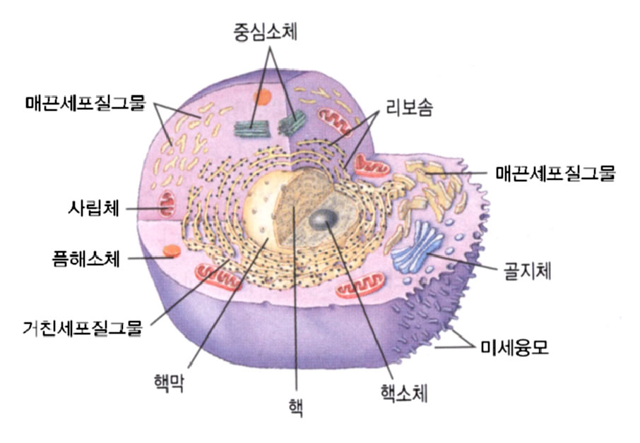 |
||
| 영문 | cell-mediated immunity | 한글 | 세포매개면역 |
|---|---|---|---|
| 설명 | 면역이란 신체를 외부의 물질로부터 보호하는 행위를 말한다. 여기에는 특이적 면역과 비특이적 면역의 두 가지가 있다. 비특이적 면역이라함은 특정한 물질에 관계하는 면역이 아니라 특정 대상이 없이 모든 외부 물체에 작용할 수 있는 면역을 말한다. 여기에는 소변의 흐름, 눈물의 흐름, 피부의 비투과성 등의 기계적인 것도 포함되고 피속에 돌아다니는 세포 중에서 비특이적으로 외부의 물질을 포식하는 세포들(예를 들면 큰포식세포(macrophage)의 활동도 포함이 된다. 세포매개면역이란 특이한 물질을 감지할 수 있는 세포를 생성하게 하여 그것으로 하여금 그 물질을 포식하게 하는 것을 말한다. |
||
| MEN | Multiple Endocrine Neoplasia ; AD Trait 1. MEN Type I(= Wermer Syndro... |
|---|---|
| MC | mass casualties; mast cell; Master of Surgery [Lat. Magister Chirurgiae]; maximum concentration; Med... |
| ICA | 1) Islet Cell Antibody 2) Internal Carotid Artery |
| IC | icteric, icterus; immune complex; immunoconjugate; immunocytochemistry; immunocytotoxicity; impedanc... |
| ICA | Institute of Clinical Analysis; internal carotid artery; intracranial aneurysm; islet cell antibody |
| ICA | Islet Cell-Cytoplasmic Antibodies |
|---|---|
| ICA | Islet cell antibodies |
| ICAs | Islet cell antibodies |
| ICSA | Islet cell surface antibodies |
| ICA | Islet cell |
| islet cell antibodies | In first-degree relatives of probands with insulin-dependent diabetes mellitus the presence of high titre ICA of the IgG cytoplasmic variety (IgG -ICA) and ICA of the complement-fixing subgroup (CF-ICA) confer a relative risk of 75 for development of insulin-dependent diabetes mellitus. The presence of ICA combined with a decrease in the first-phase of insulin secretion ( less than 25 micro U/mL) is predictive with a 95% likelihood of the development of insulin-dependent diabetes mellitus within 12 months. Reproducible results among laboratories are possible with careful attention to selection of the human pancreas as substrate as well as to the use of dilutions to generate standard curves and to the conversion of results to units. The prozone phenomena described elsewhere are not common in our experience. Fifty percent of relatives with a single positive ICA test will develop insulin-dependent diabetes mellitus within 10 years, and 60-80% of relatives with both ICA and insulin autoantibodies (IAA) will develop insulin-dependent diabetes mellitus within 10 years. The predictive value for health of negative results for ICA and IAA is almost 99%. Strong, persistently positive ICA (i.e., 40 JDF U or greater), especially if accompanied by markedly decreased insulin secretion, are the best predictors of subsequent development of insulin-dependent diabetes mellitus. The 64 kD beta-cell autoantigen long thought to be an important target for ICA is not yet available from expression cloning despite efforts by several groups.13 ICA positivity correlates with rapid loss of C-peptide secretory capacity in newly diagnosed ICA-positive insulin-dependent diabetes mellitus. The predictive values of ICA for development of insulin-dependent diabetes mellitus within 10 years in first-degree relatives of patients with insulin-dependent diabetes mellitus increase from 40% at low levels of ICA to 100% at high levels, whereas the sensitivity is 88% at low levels and 31% at high levels. In general, the risk of insulin-dependent diabetes mellitus in relatives of probands increases with titre of ICA, is greater in multiplex families, and is increased in those less than 10 years of age with positive ICA. Although the prevalence of ICA in Japanese with autoimmune thyroid disease resembles that in Caucasians, the incidence of insulin-dependent diabetes mellitus in the Japanese population is only 1/30 - 1/50 that in Caucasians. Prediabetics positive for ICA and IAA have increased suppressor-inducer (CD45R) and decreased helper-inducer (CDw29) peripheral blood lymphocytes. In two randomised, prospective, placebo-controlled studies of recent-onset insulin-dependent diabetes mellitus, cyclosporine immunosuppression increased the rate of non-insulin-requiring remissions as well as beta-cell function during drug treatment. Although 12 months of cyclosporine therapy decreases titres of ICA and insulin antibodies (IA) and increases glucagon-stimulated levels of serum C-peptide, the determination of ICA and IA and HLA-DR type are of no predictive value in selecting recent- onset insulin-dependent diabetes mellitus. Patients for cyclosporine immunointervention. Genetic control of autoimmunity in insulin-dependent diabetes mellitus is reviewed. EIA for autoantibodies to a 64 kD islet-cell protein is promising for prediction of insulin-dependent diabetes mellitus, but sensitivity and specificity are still suboptimal. See also: insulin antibodies. (05 Mar 2000) |
|---|
| islet-cell-stimulating antibodies | <immunology> Autoantibodies to a putative beta-cell receptor; stimulate the release of insulin both in rodents and man; may be analogous to the thyroid stimulating antibodies that cause grave's hyperthyroidism Synonym: icsta (05 Dec 1998) |
|---|---|
| adenoma, islet cell | A benign tumour of the islets of langerhans that may occur anywhere throughout the pancreas. Such tumours may result in hyperinsulinism or zollinger-ellison syndrome. (12 Dec 1998) |
| carcinoma, islet cell | A carcinoma of the islets of langerhans. (12 Dec 1998) |
| pancreatic isleT-cell tumours | <radiology> Insulinoma (beta-cell), usually solitary, 85% benign, gastrinoma, small, slow-growing, multiple, 60% malignant, Zollinger-Ellison syndrome: mult. Intractable ulcers, VIPoma, vasoactive intestinal peptide (VIP), WDHA syndrome: watery diarrhoea, hypokalaemia, achlorhydria, Verner-Morrison syndrome, glucagonoma, hyperglycaemia, migratory necrolytic erythema APUDomas, associated with MEN-1 (12 Dec 1998) |
| islet cell | <pathology> Cells of the Islets of Langerhans within the pancreas. See: A cells, B-cells, D cells. (18 Nov 1997) |
| islet cell adenoma | <tumour> A benign neoplasm of the pancreas composed of tissue similar in structure to that of the islets of Langerhans; it may contain functioning beta cells, and may cause hypoglycaemia. See: insulinoma. Synonym: nesidioblastoma. (05 Mar 2000) |
| islet cell antigen-related protein-tyrosine phosphatase | <enzyme> Receptor-like autoantigen found in insulin-dependent diabetes; genbank l76258 Registry number: EC 3.1.3.- Synonym: iar ptp (26 Jun 1999) |
| blood islet | An aggregation of splanchnic mesodermal cells on the embryonic yolk sac, with the potentiality of forming vascular endothelium and primitive blood cells. Synonym: blood islet. (05 Mar 2000) |
| islet | A small island. (05 Mar 2000) |
| islet amyloid peptide | <hormone, protein> Peptide of 37 amino acids that selectively inhibits insulin stimulated glucose uptake in muscle. Structurally related to calcitonin gene-related peptide. (15 Oct 1997) |
| islet neogenesis-associated protein | <chemical> Constituent of ilotropin; stimulates proliferation of pancreatic ductal cells in vitro; from hamsters and humans; amino acid sequence given in first source Synonym: ingap protein, ingap gene product (05 Dec 1998) |
| islet tissue | <anatomy> Groups of cells found within the pancreas: A cells and B-cells secrete insulin and glucagon. See: D cells. (18 Nov 1997) |
| acetylcholine receptor antibodies | <neurology, investigation> A test used to measure the amount of antibodies to acetylcholine receptors on nerve endings. This is a diagnostic test for myasthenia gravis. A normal value is no antibodies in the bloodstream. Acetylcholine receptor (AChR) binding autoantibodies (i.e. Antibodies reactive with several epitopes other than the binding site for acetylcholine or alpha-bungarotoxin) are present in approximately 88% of patients with generalised myasthenia gravis, 70% of ocular myasthenia and in approximately 80% of myasthenia gravis in remission. Although serum concentrations of AChR binding autoantibodies do not in general correlate well with severity of weakness, there is typical decrease in concentration as weakness improves with immunosuppressive therapy. AChR blocking autoantibodies (i.e., antibodies reactive with the AChR binding site) are present in about 50% of patients with myasthenia gravis, 30% with ocular myasthenia gravis and 20% of myasthenia gravis in remission, AChR blocking autoantibodies are the only AChR autoantibodies present in about 1% of myasthenia gravis. AChR modulating autoantibodies (i.e., autoantibodies which cross-link AChRs and cause their removal from muscle membrane surfaces) are present in more than 90% of myasthenia gravis and occasionally are the only AchR autoantibodies detectable in mild, recent onset or ocular-restricted myasthenia gravis. Results for AChR modulating autoantibodies can be transiently false-positive due to curare-like drugs used during general anesthesia. AChR autoantibodies of one or more types are found in at least 80% of ocular myasthenia gravis. Although generally absent in neurological conditions other than myasthenia gravis(and consequently unlikely to cause confusion in neurodiagnosis), false-positive results for AChR autoantibodies occasionally occur in primary biliary cirrhosis, tardive dyskinesia, autoimmune thyroiditis, the elderly, amyotrophic lateral sclerosis patients treated with cobra venom and patients with thymoma in the absence of myasthenia gravis. Approximately 1% of patients with rheumatoid arthritis treated with D-penicillamine develop AChR autoantibodies and myasthenia gravis, both of which disappear when the drug is discontinued. Babies born to ~10% of myasthenia gravis mothers have a transient neonatal form of myasthenia gravis that responds well to anticholinesterase therapy and usually remits within 1 month as maternal IgG disappears. (29 Dec 1997) |
| antibodies | Any of numerous protein molecules produced by the B-cells as a primary immune defense. (16 Dec 1997) |
| antibodies, anticardiolipin | Antiphospholipid antibodies found in association with systemic lupus erythematosus (lupus erythematosus, systemic), antiphospholipid syndrome, and in a variety of other diseases as well as in healthy individuals. The antibodies are detected by solid-phase immunoassay employing the purified phospholipid antigen cardiolipin. (12 Dec 1998) |
제품명 |
판매사 |
보험코드 | 성분/함량 | 구분/보험급여 |
|---|
제품명 |
판매사 |
보험코드 | 성분/함량 | 구분/보험급여 |
|---|