| 영문 | muscle cell(=muscle fiber) | 한글 | 근육세포 |
|---|---|---|---|
| 설명 | 근육은 수의근(의식에 의해서 조절이 가능한 근육: 예를 들어 다리, 팔, 얼굴근육 등)과 불수의근(의식과 무관하게 조절하지 않아도 움직이는 근육: 예를 들어 심장근, 소화기관에 분포하는 근육 등)으로 나누어지며, 근육세포의 모양은 다음과 같다(수의근, 불수의근 외에 심장에 있는 심장근은 불수의근에 해당하지만 그 모양은 수의근과 같아 따로 분류한다).  |
||
| 영문 | white blood cell(WBC), leukocyte | 한글 | 백혈구 |
|---|---|---|---|
| 설명 | 혈액내에 골수구계세포와 림프계세포, 단핵구계세포를 모두 통틀어 말한다. 백혈구의 증가가 있으면 대개 감염이 있거나, 혹은 탈수현상이 있음을 의미한다. 또한 지나친 백혈구수의 감소는 인체내 면역기능이 떨어져 있음을 의미하며, 다른 질병에 의해 나타나는 이차적인 현상이 아닌지 꼭 진단을 받아보아야 한다. |
||
| 영문 | mast cell | 한글 | 비만 세포 |
|---|---|---|---|
| 설명 | 동물의 결합 조직 가운데 널리 분포하는 세포. 결합조직과 점막조직 내에 있는 호염기성 색소로 이염색성(metachromasia)을 나타내는 과립을 가진 방추형의 세포에 작은 둥근 핵을 가진다. 비만세포의 표면에는 IgE에 대한 수용체가 존재하며, 수용체에 결합한 IgE 분자들끼리 다가의 항원에 의해 서로 연결되면 비만세포 과립탈출 반응이 일어나, 히스타민, 세로토닌, 헤파린 등의 화학전달 물질이 방출되어, 즉시형 알레르기 반응 등의 증상을 일으킨다. 피부, 장막, 혈관 주위, 점막 주변에 있다. |
||
| 영문 | cell | 한글 | 세포 |
|---|---|---|---|
| 설명 | 생명체를 이루는 구조적, 기능적 단위. 핵의 유무에 따라서 유핵세포와 무핵세포로 나눌 수가 있다. 무핵세포란 핵이 없는 세포를 말한다. 핵이란 유전정보를 가지고 있는 염색체를 보관하는 곳인데 무핵세포에서는 염색체가 세포속에 그냥 노출되어 있다. 대개 세포 하나하나가 독립된 생명체의 역할을 하는 단세포생물로서 외부환경으로부터 자신을 보호할 수 있는 견고한 세포벽을 가지고 있다. 유핵세포란 핵을 가지고 있어서 유전정보가 있는 염색체가 세포의 다른 부분과 구분되어 핵속에 들어있다. 세포속에서 핵외의 부분(이를 세포질이라 한다)에는 여러 가지 세포의 소기관이 있어서 세포의 다양한 기능을 분담한다. ◈유핵세포의 기본 구조 1.세포막-세포를 둘러싸서 주위환경과 분리하여 세포의 내환경을 유지한다. 2.형질내세망(endoplasmic reticulum)-단위막으로 둘러쌓여 있는 불규칙한 망상구조이다. 이 망상구조의 내부를 소강, 소조라고 부른다. 여기에는 표면이 매끈한 무과립형질내세망과 표면이 불규칙한 과립형질내세망의 두 가지가 있다. 과립형질내세망의 경우는 표면에 단백질을 합성하는 리보솜이라는 것이 부착되어 있다. 이곳은 주로 세포밖으로 분비할 단백질을 합성하는 장소이다. 무과립형질내세망은 표면에 리보솜이 부착되어 있지 않은 것을 말하며, 이곳에서는 주로 해독작용, 글리코겐의 합성, 스테로이드호르몬의 합성 등이 일어난다. 3.리보솜-단백질을 합성하는 역할을 하는 곳이다.이것은 세포질에 존재하는 자유리보솜과 과립형질내세망에 부착이 되어 존재하는 부착리보솜의 두 가지로 나눈다. 자유리보솜은 주로 세포내에서 필요한 단백질을 만드는 역할을 하고 부착리보솜은 세포밖으로 분비할 단백질을 만드는 역할을 한다. 4.골지장치(Golgi apparatus)-핵주위에 분포하는 납작해진 주머니모양의 것이 중첩되어 형성된 층판 모양의 구조물로 과립형질내세망에서 생성되어 외부로 분비될 단백질을 모아서 농축, 포장하여 과립을 만드는 역할을 한다. 5.사립체(mitochondria)-구형, 난형의 긴 막대기 모양으로 크기가 다양한 구조물. 생물체의 에너지 저장물질인 ATP를 생산하는 역할을 한다. 또 세포와 다른 자신만의 유전정보를 가진 DNA, RNA를 가지고 있다. 모양, 크기가 세균과 비슷하며 자체증식성 등 독립된 생명체로서 필요한 요건을 갖추고 있어서 세포와 공생관계를 가진 독립된 세포로 생각하고 있다. 6.용해소체(lysosome)-작은 구형의 소체로 여러 가지 분해효소를 가진다. 세포외계에서 들어온 물질과 결합하여 그 물질들을 용해하는 역할을 하고, 오래된 세포소기관들을 제거하는 역할도 한다. 7.세포핵(nucleus)-구형, 난형으로 세포의 중심에 위치한다. 핵내에는 유전정보가 있는 물질인 염색체가 존재한다. 8.중심소체(centrosome)-핵주위에 존재하면서 핵의 분열시에 양쪽의 염색체를 당기는 작은 섬유를 만드는 역할을 하는 곳. 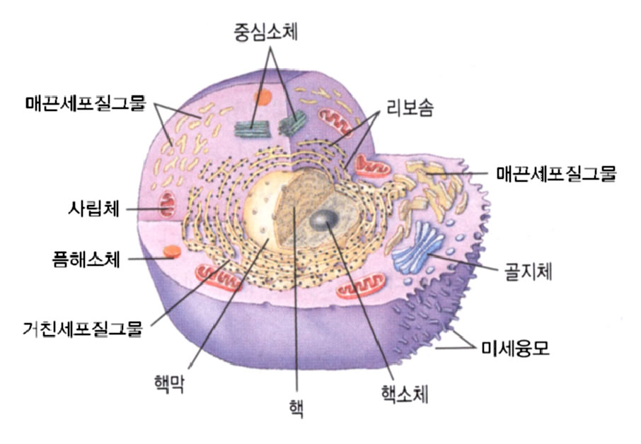 |
||
| 영문 | cell-mediated immunity | 한글 | 세포매개면역 |
|---|---|---|---|
| 설명 | 면역이란 신체를 외부의 물질로부터 보호하는 행위를 말한다. 여기에는 특이적 면역과 비특이적 면역의 두 가지가 있다. 비특이적 면역이라함은 특정한 물질에 관계하는 면역이 아니라 특정 대상이 없이 모든 외부 물체에 작용할 수 있는 면역을 말한다. 여기에는 소변의 흐름, 눈물의 흐름, 피부의 비투과성 등의 기계적인 것도 포함되고 피속에 돌아다니는 세포 중에서 비특이적으로 외부의 물질을 포식하는 세포들(예를 들면 큰포식세포(macrophage)의 활동도 포함이 된다. 세포매개면역이란 특이한 물질을 감지할 수 있는 세포를 생성하게 하여 그것으로 하여금 그 물질을 포식하게 하는 것을 말한다. |
||
| FACS | Fellow of the American College of Surgeons; fluorescence-activated cell sorter |
|---|---|
| MC | mass casualties; mast cell; Master of Surgery [Lat. Magister Chirurgiae]; maximum concentration; Med... |
| MAP | malignant atrophic papulosis; mandibular angle plane; maturation-activated protein; maximal aerobic ... |
| ACC | accommodation; acetyl coenzyme A carboxylase; acinic cell carcinoma; acute care center; adenoid cyst... |
| GC | ganglion cell; gas chromatography; general circulation; general closure; general condition; generali... |
| FACS | Fluorescence Activated Cell Sorter |
|---|---|
| FACS | Fluorescent Activated Cell Sorter |
| FACS | Fluorescence Activated Cell Sorting |
| LAK cell | lymphokine activated killer cell |
| ALCAM | Activated leukocyte cell adhesion molecule |
| fluorescence-activated cell sorter | <technique> Flow cytometry is an emerging technique which holds great promise for the separation, classification and quantitation of blood cells and antibodies which affect blood cells. Complex computerised instruments are used to pass a monocellular stream of cells, platelets or other microscopic particulate elements through a beam of laser light. The cells are categorised first by size and then computer analysed to sort the mixture of cellular elements into cell type by size. Cells are labelled with fluorescent dye and then passed, in suspending medium, through a narrow dropping nozzle so that each cell is in a small droplet. A laser based detector system is used to excite fluorescence and droplets with positively fluorescent cells are given an electric charge. Charged and uncharged droplets are separated as they fall between charged plates and so collect in different tubes. The machine can be used either as an analytical tool, counting the number of labelled cells in a population or to separate the cells for subsequent growth of the selected population. Further sophistication can be built into the system by using a second laser system at right angles to the first to look at a second fluorescent label or to gauge cell size on the basis of light scatter. The great strength of the system is that it looks at large numbers of individual cells and makes possible the separation of populations with, for example: particular surface properties. Tabulation of counted data in conjunction with size analysis enables determination of relative percentages of each specific cellular subset for which monoclonal antibody conjugates are utilised, even when the size of the cell is identical to other subset species. Flow cytometry is a slightly imprecise but common term for the use of the Fluorescence-activated Cell Sorter (FACS). (01 Dec 1998) |
|---|---|
| fluorescence-activated cell sorting | <technique> A technique for separating and sorting cells marked with a fluorescent label based on how much they fluoresce at a particular wavelength. (12 Jan 1998) |
| cell sorter | <apparatus> A device used to separate different kinds of cells from a mixed, or heterogeneous, population. (26 Mar 1998) |
| wool-sorter's disease | A form of anthrax acquired by inhalation of dust containing Bacillus anthracis; there is an initial chill followed by pain in the back and legs, rapid respiration, dyspnea, cough, fever, rapid pulse, and extreme cardiovascular collapse. Synonym: anthrax pneumonia, ragpicker's disease, ragsorter's disease, rag-sorter's disease, wool-sorter's pneumonia, woolsorter's disease, wool-sorter's disease. (05 Mar 2000) |
| wool-sorter's pneumonia | A form of anthrax acquired by inhalation of dust containing Bacillus anthracis; there is an initial chill followed by pain in the back and legs, rapid respiration, dyspnea, cough, fever, rapid pulse, and extreme cardiovascular collapse. Synonym: anthrax pneumonia, ragpicker's disease, ragsorter's disease, rag-sorter's disease, wool-sorter's pneumonia, woolsorter's disease, wool-sorter's disease. (05 Mar 2000) |
| platelet endothelial cell activated protease | <enzyme> Degrades casein and fibrinogen; secreted by endothelial cells and activated in the extracellular medium by platelets; not inhibited by serine protease inhibitors, metalloproteinase inhibitors, or cystein protease inhibitors; pH optimum 7.5 Registry number: EC 3.4.99.- Synonym: pecap (26 Jun 1999) |
| immunologically activated cell | An immunocyte that is in an elevated state of reactivity capable of carrying out an immune response, in contradistinction to an immunologically competent cell. (05 Mar 2000) |
| ratio imaging fluorescence microscopy | <procedure> A method of measurement of intracellular pH or intracellular calcium levels, using a fluorescent probe molecule (see fura-2), in which the two different excitation wavelengths are used and the emitted light levels compared. If emission at one wavelength is sensitive to the intracellular ion level and emission at the other wavelength is not, then standardisation for intracellular probe concentration, efficiency of light collection, inactivation of probe and thickness of cytoplasm can all be performed automatically. (17 Dec 1997) |
| microscopy, fluorescence | Microscopy of specimens stained with fluorescent dye (usually fluorescein isothiocyanate) or of naturally fluorescent materials, which emit light when exposed to ultraviolet or blue light. Immunofluorescence microscopy utilises antibodies that are labelled with fluorescent dye. (12 Dec 1998) |
| spectrometry, fluorescence | Measurement of the intensity and quality of fluorescence. (12 Dec 1998) |
| Eranko's fluorescence stain | <technique> Exposure of frozen sections to formaldehyde which produces a strong yellow-green fluorescence from cells containing norepinephrine. (05 Mar 2000) |
| fluorescence | <chemistry, physics> The emission of one or more photons by a molecule or atom activated by the absorption of a quantum of electro magnetic radiation. Typically the emission, that is of longer wavelength than the excitatory radiation, occurs within 10exp 8 seconds: phosphorescence is a phenomenon with a longer or much longer delay in re radiation. Note that rays, X-rays, UV, visible light and IR radiations may all stimulate fluorescence. (25 Jun 1999) |
| fluorescence energy transfer | <technique> Transfer of energy from one fluorochrome to another. The emission wavelength of the fluorochrome excited by the incident light must approximately match the excitation wavelength of the second fluorochrome. If light at the second emission wavelength is detected, it implies that the two fluorochromes were physically within a few nanometres. Used as a technique to probe protein or cell interactions. (25 Jun 1999) |
| fluorescence immunoassay | <technique> A sensitive technique which uses fluorescein, a fluorescent molecule, to measure the antigen or antibody concentration in a solution. (09 Oct 1997) |
| fluorescence in situ hybridization | <molecular biology, technique> A type of in situ hybridization in which target sequences are stained with fluorescent dye so their location and size can be determined using fluorescence microscopy. This staining is sufficiently distinct that the hybridization signal can be seen both in metaphase spreads and in interphase nuclei. Acronym: FISH (25 Jun 1999) |
제품명 |
판매사 |
보험코드 | 성분/함량 | 구분/보험급여 |
|---|
제품명 |
판매사 |
보험코드 | 성분/함량 | 구분/보험급여 |
|---|