| ПЕЙЎ | mast cell | ЧбБл | КёИИ ММЦї |
|---|---|---|---|
| МГИэ | ЕПЙАРЧ АсЧе СЖСї АЁПюЕЅ ГЮИЎ КаЦїЧЯДТ ММЦї. АсЧеСЖСїАњ СЁИЗСЖСї ГЛПЁ РжДТ ШЃПАБтМК ЛіМвЗЮ РЬПАЛіМК(metachromasia)РЛ ГЊХИГЛДТ АњИГРЛ АЁСј ЙцУпЧќРЧ ММЦїПЁ РлРК ЕеБй ЧйРЛ АЁСјДй. КёИИММЦїРЧ ЧЅИщПЁДТ IgEПЁ ДыЧб МіПыУМАЁ СИРчЧЯИч, МіПыУМПЁ АсЧеЧб IgE КаРкЕщГЂИЎ ДйАЁРЧ ЧзПјПЁ РЧЧи МЗЮ ПЌАсЕЧИщ КёИИММЦї АњИГХЛУт ЙнРРРЬ РЯОюГЊ, ШїНКХИЙЮ, ММЗЮХфДб, ЧьЦФИА ЕюРЧ ШЧаРќДо ЙАСњРЬ ЙцУтЕЧОю, СяНУЧќ ОЫЗЙИЃБт ЙнРР ЕюРЧ СѕЛѓРЛ РЯРИХВДй. ЧЧКЮ, РхИЗ, ЧїАќ СжРЇ, СЁИЗ СжКЏПЁ РжДй. |
||
| ПЕЙЎ | cell | ЧбБл | ММЦї |
|---|---|---|---|
| МГИэ | Л§ИэУМИІ РЬЗчДТ БИСЖРћ, БтДЩРћ ДмРЇ. ЧйРЧ РЏЙЋПЁ ЕћЖѓМ РЏЧйММЦїПЭ ЙЋЧйММЦїЗЮ ГЊД МіАЁ РжДй. ЙЋЧйММЦїЖѕ ЧйРЬ ОјДТ ММЦїИІ ИЛЧбДй. ЧйРЬЖѕ РЏРќСЄКИИІ АЁСіАэ РжДТ ПАЛіУМИІ КИАќЧЯДТ АїРЮЕЅ ЙЋЧйММЦїПЁМДТ ПАЛіУМАЁ ММЦїМгПЁ БзГЩ ГыУтЕЧОю РжДй. ДыАГ ММЦї ЧЯГЊЧЯГЊАЁ ЕЖИГЕШ Л§ИэУМРЧ ПЊЧвРЛ ЧЯДТ ДмММЦїЛ§ЙАЗЮМ ПмКЮШЏАцРИЗЮКЮХЭ РкНХРЛ КИШЃЧв Мі РжДТ АпАэЧб ММЦїКЎРЛ АЁСіАэ РжДй. РЏЧйММЦїЖѕ ЧйРЛ АЁСіАэ РжОюМ РЏРќСЄКИАЁ РжДТ ПАЛіУМАЁ ММЦїРЧ ДйИЅ КЮКаАњ БИКаЕЧОю ЧйМгПЁ ЕщОюРжДй. ММЦїМгПЁМ ЧйПмРЧ КЮКа(РЬИІ ММЦїСњРЬЖѓ ЧбДй)ПЁДТ ПЉЗЏ АЁСі ММЦїРЧ МвБтАќРЬ РжОюМ ММЦїРЧ ДйОчЧб БтДЩРЛ КаДуЧбДй. ЂТРЏЧйММЦїРЧ БтКЛ БИСЖ 1.ММЦїИЗ-ММЦїИІ ЕбЗЏНЮМ СжРЇШЏАцАњ КаИЎЧЯПЉ ММЦїРЧ ГЛШЏАцРЛ РЏСіЧбДй. 2.ЧќСњГЛММИС(endoplasmic reticulum)-ДмРЇИЗРИЗЮ ЕбЗЏНзПЉ РжДТ КвБдФЂЧб ИСЛѓБИСЖРЬДй. РЬ ИСЛѓБИСЖРЧ ГЛКЮИІ МвА, МвСЖЖѓАэ КЮИЅДй. ПЉБтПЁДТ ЧЅИщРЬ ИХВіЧб ЙЋАњИГЧќСњГЛММИСАњ ЧЅИщРЬ КвБдФЂЧб АњИГЧќСњГЛММИСРЧ ЕЮ АЁСіАЁ РжДй. АњИГЧќСњГЛММИСРЧ АцПьДТ ЧЅИщПЁ ДмЙщСњРЛ ЧеМКЧЯДТ ИЎКИМиРЬЖѓДТ АЭРЬ КЮТјЕЧОю РжДй. РЬАїРК СжЗЮ ММЦїЙлРИЗЮ КаКёЧв ДмЙщСњРЛ ЧеМКЧЯДТ РхМвРЬДй. ЙЋАњИГЧќСњГЛММИСРК ЧЅИщПЁ ИЎКИМиРЬ КЮТјЕЧОю РжСі ОЪРК АЭРЛ ИЛЧЯИч, РЬАїПЁМДТ СжЗЮ ЧиЕЖРлПы, БлИЎФкАеРЧ ЧеМК, НКХзЗЮРЬЕхШЃИЃИѓРЧ ЧеМК ЕюРЬ РЯОюГДй. 3.ИЎКИМи-ДмЙщСњРЛ ЧеМКЧЯДТ ПЊЧвРЛ ЧЯДТ АїРЬДй.РЬАЭРК ММЦїСњПЁ СИРчЧЯДТ РкРЏИЎКИМиАњ АњИГЧќСњГЛММИСПЁ КЮТјРЬ ЕЧОю СИРчЧЯДТ КЮТјИЎКИМиРЧ ЕЮ АЁСіЗЮ ГЊДЋДй. РкРЏИЎКИМиРК СжЗЮ ММЦїГЛПЁМ ЧЪПфЧб ДмЙщСњРЛ ИИЕхДТ ПЊЧвРЛ ЧЯАэ КЮТјИЎКИМиРК ММЦїЙлРИЗЮ КаКёЧв ДмЙщСњРЛ ИИЕхДТ ПЊЧвРЛ ЧбДй. 4.АёСіРхФЁ(Golgi apparatus)-ЧйСжРЇПЁ КаЦїЧЯДТ ГГРлЧиСј СжИгДЯИ№ОчРЧ АЭРЬ СпУИЕЧОю ЧќМКЕШ УўЦЧ И№ОчРЧ БИСЖЙАЗЮ АњИГЧќСњГЛММИСПЁМ Л§МКЕЧОю ПмКЮЗЮ КаКёЕЩ ДмЙщСњРЛ И№ОЦМ ГѓУр, ЦїРхЧЯПЉ АњИГРЛ ИИЕхДТ ПЊЧвРЛ ЧбДй. 5.ЛчИГУМ(mitochondria)-БИЧќ, ГЧќРЧ Бф ИЗДыБт И№ОчРИЗЮ ХЉБтАЁ ДйОчЧб БИСЖЙА. Л§ЙАУМРЧ ПЁГЪСі РњРхЙАСњРЮ ATPИІ Л§ЛъЧЯДТ ПЊЧвРЛ ЧбДй. ЖЧ ММЦїПЭ ДйИЅ РкНХИИРЧ РЏРќСЄКИИІ АЁСј DNA, RNAИІ АЁСіАэ РжДй. И№Оч, ХЉБтАЁ ММБеАњ КёНСЧЯИч РкУМСѕНФМК Ею ЕЖИГЕШ Л§ИэУМЗЮМ ЧЪПфЧб ПфАЧРЛ АЎУпАэ РжОюМ ММЦїПЭ АјЛ§АќАшИІ АЁСј ЕЖИГЕШ ММЦїЗЮ Л§АЂЧЯАэ РжДй. 6.ПыЧиМвУМ(lysosome)-РлРК БИЧќРЧ МвУМЗЮ ПЉЗЏ АЁСі КаЧиШПМвИІ АЁСјДй. ММЦїПмАшПЁМ ЕщОюПТ ЙАСњАњ АсЧеЧЯПЉ Бз ЙАСњЕщРЛ ПыЧиЧЯДТ ПЊЧвРЛ ЧЯАэ, ПРЗЁЕШ ММЦїМвБтАќЕщРЛ СІАХЧЯДТ ПЊЧвЕЕ ЧбДй. 7.ММЦїЧй(nucleus)-БИЧќ, ГЧќРИЗЮ ММЦїРЧ СпНЩПЁ РЇФЁЧбДй. ЧйГЛПЁДТ РЏРќСЄКИАЁ РжДТ ЙАСњРЮ ПАЛіУМАЁ СИРчЧбДй. 8.СпНЩМвУМ(centrosome)-ЧйСжРЇПЁ СИРчЧЯИщМ ЧйРЧ КаПНУПЁ ОчТЪРЧ ПАЛіУМИІ ДчБтДТ РлРК МЖРЏИІ ИИЕхДТ ПЊЧвРЛ ЧЯДТ Аї. 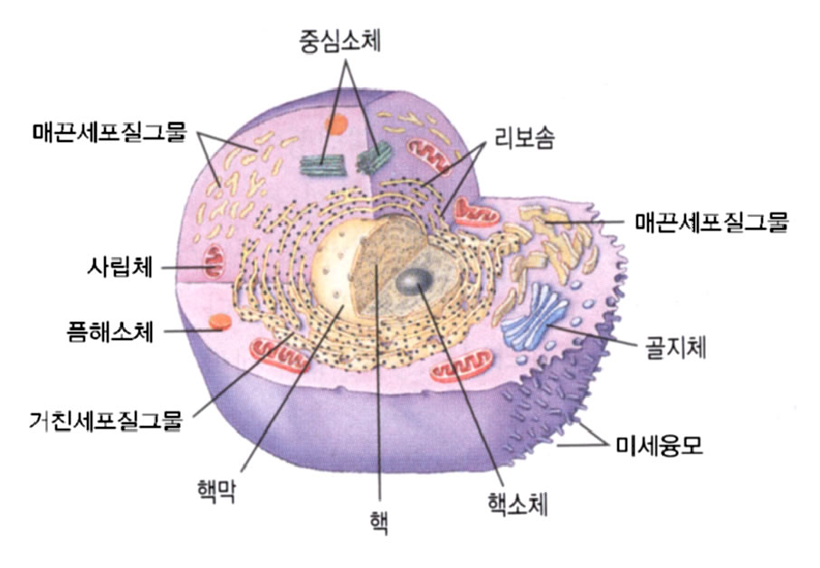 |
||
| ПЕЙЎ | cell-mediated immunity | ЧбБл | ММЦїИХАГИщПЊ |
|---|---|---|---|
| МГИэ | ИщПЊРЬЖѕ НХУМИІ ПмКЮРЧ ЙАСњЗЮКЮХЭ КИШЃЧЯДТ ЧрРЇИІ ИЛЧбДй. ПЉБтПЁДТ ЦЏРЬРћ ИщПЊАњ КёЦЏРЬРћ ИщПЊРЧ ЕЮ АЁСіАЁ РжДй. КёЦЏРЬРћ ИщПЊРЬЖѓЧдРК ЦЏСЄЧб ЙАСњПЁ АќАшЧЯДТ ИщПЊРЬ ОЦДЯЖѓ ЦЏСЄ ДыЛѓРЬ ОјРЬ И№Еч ПмКЮ ЙАУМПЁ РлПыЧв Мі РжДТ ИщПЊРЛ ИЛЧбДй. ПЉБтПЁДТ МвКЏРЧ ШхИЇ, ДЋЙАРЧ ШхИЇ, ЧЧКЮРЧ КёХѕАњМК ЕюРЧ БтАшРћРЮ АЭЕЕ ЦїЧдЕЧАэ ЧЧМгПЁ ЕЙОЦДйДЯДТ ММЦї СпПЁМ КёЦЏРЬРћРИЗЮ ПмКЮРЧ ЙАСњРЛ ЦїНФЧЯДТ ММЦїЕщ(ПЙИІ ЕщИщ ХЋЦїНФММЦї(macrophage)РЧ ШАЕПЕЕ ЦїЧдРЬ ЕШДй. ММЦїИХАГИщПЊРЬЖѕ ЦЏРЬЧб ЙАСњРЛ АЈСіЧв Мі РжДТ ММЦїИІ Л§МКЧЯАд ЧЯПЉ БзАЭРИЗЮ ЧЯПЉБн Бз ЙАСњРЛ ЦїНФЧЯАд ЧЯДТ АЭРЛ ИЛЧбДй. |
||
| ПЕЙЎ | nerve cell | ЧбБл | НХАцММЦї |
|---|---|---|---|
| МГИэ | НХАцММЦїДТ ПУЙйИЅ НХАцРќДоРЛ РЇЧб АЂ КЮКаКАЗЮ ГЊДЕОюСЎ РжДй. НХАцММЦїПЁМДТ РќЧиСЎПРДТ РкБиРЛ РќБтРћРЮ НХШЃЗЮ ЙйВюОю КИГЛАХГЊ ЙоАд ЕШДй. РЬЗБ РќБтРћРЮ ЧіЛѓРК АЂ НХАцММЦїГЛПЁ СИРчЧЯДТ АЂ РЬПТУЄГЮ(ion channel: ionРЬЖѕ ГЊЦЎЗ§, ФЎЗ§ ЕюРЛ СіФЊЧЯДТ ИЛЕщЗЮНс, РЬЕщРЬ ММЦїИЗПЁ РЧЧи ГЊДЕОюСњ ЖЇ Л§БтДТ РќОаТїАЁ РќБтРћ РкБиРЛ РЯРИХААэ РЏСіЧЯДТЕЅ АсСЄРћРЮ ПЊЧвРЛ ЧбДй)ЕщРЧ РлПыПЁ РЧЧи РЬЗчОюСіАд ЕШДй. |
||
| ПЕЙЎ | glia cell | ЧбБл | ОЦБГММЦї |
|---|---|---|---|
| МГИэ | НХАцММЦї ЛчРЬПЁМ БзЙАБИСЖИІ РЬЗчИч РЬИІ СіСіЧЯДТ СЖСї. НХАцОЦБГММЦїДТ НХАцИ№ММЦїПЭ АЅЖѓСј ОЦБГИ№ММЦїАЁ ДйНУ ПЉЗЏ ЧќХТЗЮ КаШ-МКРхЧб АЭРЬДй. ГњНЧРЬГЊ УДМіСпНЩАќРЧ КЎРЛ ЕЄАэ ПјСжЛѓ ЖЧДТ РдЙцЧќРЬИч, УЪБтПЁДТ РЏИЎИщПЁ МЖИ№АЁ РжДй. ДыЧќММЦїДТ КАГњНЧИЗММЦїДТ ОЦБГММЦїЖѓАэ ЧЯИч, НХАцММЦїГЊ НХАцМЖРЏ ЛчРЬПЁ ЛъРчЧбДй. Бз ПмПЁ ШёМвЕЙБтОЦБГММЦїЕЕ ЦїЧдЕШДй. |
||
| MCC | mean corpuscular hemoglobin concentration; medial cell column; Medical Council of Canada; metacerebr... |
|---|---|
| RGC | radio-gas chromatography; remnant gastric cancer; retinal ganglion cell; right giant cell |
| GC | ganglion cell; gas chromatography; general circulation; general closure; general condition; generali... |
| TCC | Transitional Cell Cancer |
| CAR | Canadian Association of Radiologists; cancer-associated retinopathy; cardiac ambulation routine; cel... |
| cancer en cuirasse | A carcinoma that involves a considerable portion of the skin of one or both sides of the thorax. Origin: Fr. Breastplate (05 Mar 2000) |
|---|---|
| cancer family | A group of blood relatives of whom several have had cancer; the mode of aggregation may be genetic and homogeneous, as in familial polyposis of the colon; diverse as in neurofibromatosis; or due to common exposure to a carcinogenic or oncogenic agent, such as a virus. (05 Mar 2000) |
| cancer, gastric | Cancer of the stomach, the major organ that holds food for digestion. Stomach cancer (gastric cancer) can develop in any part of the stomach and spread to other organs. Stomach ulcers do not appear to increase a person's risk of developing stomach cancer. Symptoms of stomach cancer are often vague, such as loss of appetite and weight. The cancer is diagnosed with a biopsy of stomach tissue during a procedure called an endoscopy. (12 Dec 1998) |
| cancer, hodgkin's disease | A type of lymphoma (cancer of the lymphatic system). The most common symptom of Hodgkin's disease is a painless swelling in the lymph nodes in the neck, underarm, or groin. Hodgkin's disease is diagnosed when abnormal tissue is detected by a pathologist after a biopsy of an enlarged lymph node. Treatment usually includes radiation therapy or chemotherapy. Regular follow-up examinations are important after treatment for Hodgkin's disease. Patients treated for Hodgkin's disease have an increased risk of developing other types of cancer later in life, especially leukaemia. (12 Dec 1998) |
| cancer juice | Turbid, white to yellow-white or gray-white fluid (chiefly plasma) that may be expressed from certain forms of malignant neoplastic tissue, and is likely to contain neoplastic cells and debris; formed especially in relatively large, degenerating, partly necrotic foci of rapidly growing neoplastic tissue. (05 Mar 2000) |
| cancer, kidney | Cancer of the major organ responsible for the removal from the blood of the toxins of body metabolism the kidney. Childhood kidney cancer is different from the adult kidney cancer. The most common symptom of kidney cancer is blood in the urine. The diagnosis of kidney cancer is supported by findings of the medical history and examination, blood, urine, and X-ray tests, and confirmed with a biopsy. Kidney cancer is treated with surgery, embolization, radiation therapy, hormone therapy, biological therapy, or chemotherapy. (12 Dec 1998) |
| cancer, larynx | Cancer of the voice box. The larynx is the voice box located at the top of the windpipe (trachea). Cancer of the larynx occurs most often in people over the age of 55 years. People who stop smoking can greatly reduce their risk of cancer of the larynx. Painless hoarseness can be a symptom of cancer of the larynx. The larynx can be examined with a viewing tube called a laryngoscope. Cancer of the larynx is usually treated with radiation therapy or surgery. Chemotherapy can also be used for cancers that have spread. (12 Dec 1998) |
| cancer, leukaemia | Leukaemia is a cancer of the white blood cells. Leukaemias are grouped by how quickly the disease develops (acute or chronic) as well as by the type of blood cell that is affected. People with leukaemia are at significantly increased risk for developing infections, anaemia, and bleeding. Diagnosis of leukaemia is supported by findings of the medical history and examination, and examining blood under a microscope. Leukaemia cells can be detected and further classified with a bone marrow aspiration and/or biopsy. most patients with leukaemia are treated with chemotherapy. Some patients also may have radiation therapy and/or bone marrow transplantation. (12 Dec 1998) |
| cancer, lung | Cancer of the major organ of respiration the lung. Lung cancer kills more men and women than any other form of cancer. Since the majority of lung cancer is diagnosed at a relatively late stage, only 10% of all lung cancer patients are ultimately cured. Eight out of 10 lung cancers are due to tobacco smoke. Lung cancers are classified as either small cell or non-small cell cancers. Persistent cough and bloody sputum can be symptoms of lung cancer. Lung cancer can be diagnosed based on examination of sputum, or tissue examination with biopsy using bronchoscopy, needle through the chest wall, or surgical excision. (12 Dec 1998) |
| cancer, lymphoma, hodgkin's | A type of lymphoma (cancer of the lymphatic system). The most common symptom of Hodgkin's disease is a painless swelling in the lymph nodes in the neck, underarm, or groin. Hodgkin's disease is diagnosed when abnormal tissue is detected by a pathologist after a biopsy of an enlarged lymph node. Treatment usually includes radiation therapy or chemotherapy. Regular follow-up examinations are important after treatment for Hodgkin's disease. Patients treated for Hodgkin's disease have an increased risk of developing other types of cancer later in life, especially leukaemia. (12 Dec 1998) |
| cancer, lymphoma, non-hodgkin's | A lymphoma is a cancer that develops in the lymphatic system. The most common symptom of non-Hodgkin's lymphomas is a painless swelling in the lymph nodes in the neck, underarm, or groin. Non-Hodgkin's lymphomas are diagnosed with a biopsy of an enlarged lymph node. Follow-up examinations are important after lymphoma treatment. Most relapses occur in the first 2 years after therapy. (12 Dec 1998) |
| cancer, malignant melanoma | A skin cancer that begins in cells called melanocytes that can grow together to form benign (not cancerous) moles. A change in size, shape, or colour of a mole can be a sign of melanoma. Melanoma can be cured if detected early, before spread (metastasis) to other areas of the body. Diagnosis is confirmed with a biopsy of the abnormal skin. Sun exposure can cause skin damage that can lead to melanoma. (12 Dec 1998) |
| cancer, melanoma | A skin cancer that begins in cells called melanocytes that can grow together to form benign (not cancerous) moles. A change in size, shape, or colour of a mole can be a sign of melanoma. It can be cured if detected early, before spread (metastasis) to other areas. Diagnosis is confirmed by a biopsy of the abnormal skin. Sun exposure can cause skin damage that can lead to melanoma. (12 Dec 1998) |
| cancer, multiple myeloma | A bone marrow cancer involving a type of white blood cell called a plasma (or myeloma) cell. The tumour cells can form a single collection (a plasmacytoma) or many tumours (multiple myeloma). Plasma cells are part of the immune system and make antibodies. Because patients have an excess of identical plasma cells, they have too much of one type of antibody. As myeloma cells increase in number, they damage and weaken the bones, causing pain and often fractures. When bones are damaged, calcium is released into the blood leading to hypercalcaemia (excess calcium in the blood) and that causes loss of appetite, nausea, thirst, fatigue, muscle weakness, restlessness, and confusion. Myeloma cells prevent the bone marrow from forming normal plasma cells and other white blood cells important to the immune system so patients may not be able to fight infections. The cancer cells can also prevent the growth of new red blood cells, causing anaemia. Excess antibody proteins and calcium may prevent the kidneys from filtering and cleaning the blood properly Cancer, non-Hodgkin's lymphoma: A lymphoma is a cancer that develops in the lymphatic system. The most common symptom of non-Hodgkin's lymphomas is a painless swelling in the lymph nodes in the neck, underarm, or groin. Non-Hodgkin's lymphomas are diagnosed with a biopsy of an enlarged lymph node. Follow-up examinations are important after lymphoma treatment. Most relapses occur in the first 2 years after therapy. (12 Dec 1998) |
| cancer, myeloma | A bone marrow cancer involving a type of white blood cell called a plasma (or myeloma) cell. The tumour cells can form a single collection (a plasmacytoma) or many tumours (multiple myeloma). Plasma cells are part of the immune system and make antibodies. Because patients have an excess of identical plasma cells, they have too much of one type of antibody. As myeloma cells increase in number, they damage and weaken the bones, causing pain and often fractures. When bones are damaged, calcium is released into the blood leading to hypercalcaemia (too much calcium in the blood) and that causes loss of appetite, nausea, thirst, fatigue, muscle weakness, restlessness, and confusion. Myeloma cells prevent the bone marrow from forming normal plasma cells and other white blood cells important to the immune system so patients may not be able to fight infections. The cancer cells can also prevent the growth of new red blood cells, causing anaemia. Excess antibody proteins and calcium may prevent the kidneys from filtering and cleaning the blood properly. (12 Dec 1998) |
СІЧАИэ |
ЦЧИХЛч |
КИЧшФкЕх | МККа/ЧдЗЎ | БИКа/КИЧшБоПЉ |
|---|
СІЧАИэ |
ЦЧИХЛч |
КИЧшФкЕх | МККа/ЧдЗЎ | БИКа/КИЧшБоПЉ |
|---|