¼±Åà - È­»ìǥŰ/¿£ÅÍŰ ´Ý±â - ESC

 
"squamous cell layer"¿¡ ´ëÇÑ °Ë»ö °á°úÀÔ´Ï´Ù. °Ë»ö °á°ú º¸´Â µµÁß¿¡ Tab ۸¦ ´©¸£½Ã¸é °Ë»ö âÀÌ ¼±Åõ˴ϴÙ.
¾Ë±â½¬¿î ÀÇÇпë¾îÇ®ÀÌÁý, ¼­¿ïÀÇ´ë ±³¼ö ÁöÁ¦±Ù, °í·ÁÀÇÇÐ ÃâÆÇ À¯»ç °Ë»ö °á°ú : 5 ÆäÀÌÁö: 2
¿µ¹® mast cell ÇÑ±Û ºñ¸¸ ¼¼Æ÷
¼³¸í   
  µ¿¹°ÀÇ °áÇÕ Á¶Á÷ °¡¿îµ¥ ³Î¸® ºÐÆ÷Çϴ ¼¼Æ÷. °áÇÕÁ¶Á÷°ú Á¡¸·Á¶Á÷ ³»¿¡ Àִ ȣ¿°±â¼º »ö¼Ò·Î ÀÌ¿°»ö¼º(metachromasia)À» ³ªÅ¸³»´Â °ú¸³À» °¡Áø ¹æÃßÇüÀÇ ¼¼Æ÷¿¡ ÀÛÀº µÕ±Ù ÇÙÀ» °¡Áø´Ù. ºñ¸¸¼¼Æ÷ÀǠǥ¸é¿¡´Â IgE¿¡ ´ëÇÑ ¼ö¿ëü°¡ Á¸ÀçÇϸç, ¼ö¿ëü¿¡ °áÇÕÇÑ IgE ºÐÀڵ鳢¸® ´Ù°¡ÀÇ Ç׿ø¿¡ ÀÇÇØ ¼­·Î ¿¬°áµÇ¸é ºñ¸¸¼¼Æ÷ °ú¸³Å»Ãâ ¹ÝÀÀÀÌ ÀϾ, È÷½ºÅ¸¹Î, ¼¼·ÎÅä´Ñ, ÇìÆÄ¸° µîÀÇ È­ÇÐÀü´Þ ¹°ÁúÀÌ ¹æÃâµÇ¾î, Áï½ÃÇü ¾Ë·¹¸£±â ¹ÝÀÀ µîÀÇ Áõ»óÀ» ÀÏÀ¸Å²´Ù. ÇǺÎ, À帷, Ç÷°ü ÁÖÀ§, Á¡¸· ÁÖº¯¿¡ ÀÖ´Ù.
¿µ¹® cell ÇÑ±Û ¼¼Æ÷
¼³¸í   
  »ý¸íü¸¦ ÀÌ·ç´Â ±¸Á¶Àû, ±â´ÉÀû ´ÜÀ§.
  
  ÇÙÀÇ À¯¹«¿¡ µû¶ó¼­ À¯ÇÙ¼¼Æ÷¿Í ¹«ÇÙ¼¼Æ÷·Î ³ª´­ ¼ö°¡ ÀÖ´Ù. ¹«ÇÙ¼¼Æ÷¶õ ÇÙÀÌ ¾ø´Â ¼¼Æ÷¸¦ ¸»ÇÑ´Ù. ÇÙÀ̶õ À¯ÀüÁ¤º¸¸¦ °¡Áö°í Àִ ¿°»öü¸¦ º¸°üÇϴ °÷Àε¥ ¹«ÇÙ¼¼Æ÷¿¡¼­´Â ¿°»öü°¡ ¼¼Æ÷¼Ó¿¡ ±×³É ³ëÃâµÇ¾î ÀÖ´Ù. ´ë°³ ¼¼Æ÷ ÇϳªÇϳª°¡ µ¶¸³µÈ »ý¸íüÀÇ ¿ªÇÒÀ» Çϴ ´Ü¼¼Æ÷»ý¹°·Î¼­ ¿ÜºÎȯ°æÀ¸·ÎºÎÅÍ ÀÚ½ÅÀ» º¸È£ÇÒ ¼ö Àִ °ß°íÇÑ ¼¼Æ÷º®À» °¡Áö°í ÀÖ´Ù. À¯ÇÙ¼¼Æ÷¶õ ÇÙÀ» °¡Áö°í À־ À¯ÀüÁ¤º¸°¡ Àִ ¿°»öü°¡ ¼¼Æ÷ÀÇ ´Ù¸¥ ºÎºÐ°ú ±¸ºÐµÇ¾î ÇÙ¼Ó¿¡ µé¾îÀÖ´Ù. ¼¼Æ÷¼Ó¿¡¼­ ÇÙ¿ÜÀÇ ºÎºÐ(À̸¦ ¼¼Æ÷ÁúÀ̶ó ÇÑ´Ù)¿¡´Â ¿©·¯ °¡Áö ¼¼Æ÷ÀÇ ¼Ò±â°üÀÌ À־ ¼¼Æ÷ÀÇ ´Ù¾çÇÑ ±â´ÉÀ» ºÐ´ãÇÑ´Ù.
  
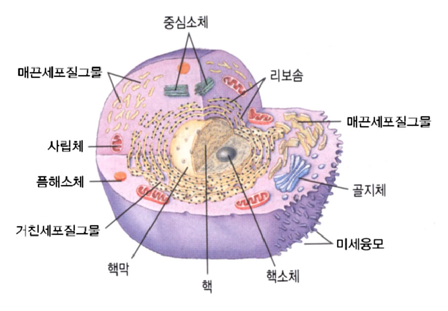
  ¢ÂÀ¯ÇÙ¼¼Æ÷ÀÇ ±âº» ±¸Á¶
  
  1.¼¼Æ÷¸·-¼¼Æ÷¸¦ µÑ·¯½Î¼­ ÁÖÀ§È¯°æ°ú ºÐ¸®ÇÏ¿© ¼¼Æ÷ÀÇ ³»È¯°æÀ» À¯ÁöÇÑ´Ù.
  
  2.ÇüÁú³»¼¼¸Á(endoplasmic reticulum)-´ÜÀ§¸·À¸·Î µÑ·¯½×¿© Àִ ºÒ±ÔÄ¢ÇÑ ¸Á»ó±¸Á¶ÀÌ´Ù. ÀÌ ¸Á»ó±¸Á¶ÀÇ ³»ºÎ¸¦ ¼Ò°­, ¼ÒÁ¶¶ó°í ºÎ¸¥´Ù. ¿©±â¿¡´Â Ç¥¸éÀÌ ¸Å²öÇÑ ¹«°ú¸³ÇüÁú³»¼¼¸Á°ú Ç¥¸éÀÌ ºÒ±ÔÄ¢ÇÑ °ú¸³ÇüÁú³»¼¼¸ÁÀÇ µÎ °¡Áö°¡ ÀÖ´Ù. °ú¸³ÇüÁú³»¼¼¸ÁÀÇ °æ¿ì´Â Ç¥¸é¿¡ ´Ü¹éÁúÀ» ÇÕ¼ºÇϴ ¸®º¸¼ØÀ̶ó´Â °ÍÀÌ ºÎÂøµÇ¾î ÀÖ´Ù. À̰÷Àº ÁַΠ¼¼Æ÷¹ÛÀ¸·Î ºÐºñÇÒ ´Ü¹éÁúÀ» ÇÕ¼ºÇϴ Àå¼ÒÀÌ´Ù. ¹«°ú¸³ÇüÁú³»¼¼¸ÁÀº Ç¥¸é¿¡ ¸®º¸¼ØÀÌ ºÎÂøµÇ¾î ÀÖÁö ¾ÊÀº °ÍÀ» ¸»Çϸç, À̰÷¿¡¼­´Â ÁÖ·Î ÇØµ¶ÀÛ¿ë, ±Û¸®ÄÚ°ÕÀÇ ÇÕ¼º, ½ºÅ×·ÎÀ̵åÈ£¸£¸óÀÇ ÇÕ¼º µîÀÌ ÀϾ´Ù.
  
  3.¸®º¸¼Ø-´Ü¹éÁúÀ» ÇÕ¼ºÇϴ ¿ªÇÒÀ» Çϴ °÷ÀÌ´Ù.À̰ÍÀº ¼¼Æ÷Áú¿¡ Á¸ÀçÇϴ ÀÚÀ¯¸®º¸¼Ø°ú °ú¸³ÇüÁú³»¼¼¸Á¿¡ ºÎÂøÀÌ µÇ¾î Á¸ÀçÇϴ ºÎÂø¸®º¸¼ØÀÇ µÎ °¡Áö·Î ³ª´«´Ù. ÀÚÀ¯¸®º¸¼ØÀº ÁַΠ¼¼Æ÷³»¿¡¼­ ÇÊ¿äÇÑ ´Ü¹éÁúÀ» ¸¸µå´Â ¿ªÇÒÀ» Ç졒ʼÎÂø¸®º¸¼ØÀº ¼¼Æ÷¹ÛÀ¸·Î ºÐºñÇÒ ´Ü¹éÁúÀ» ¸¸µå´Â ¿ªÇÒÀ» ÇÑ´Ù.
  
  4.°ñÁöÀåÄ¡(Golgi apparatus)-ÇÙÁÖÀ§¿¡ ºÐÆ÷Çϴ ³³ÀÛÇØÁø ÁָӴϸð¾çÀÇ °ÍÀÌ ÁßøµÇ¾î Çü¼ºµÈ ÃþÆÇ ¸ð¾çÀÇ ±¸Á¶¹°·Î °ú¸³ÇüÁú³»¼¼¸Á¿¡¼­ »ý¼ºµÇ¾î ¿ÜºÎ·Î ºÐºñµÉ ´Ü¹éÁúÀ» ¸ð¾Æ¼­ ³óÃà, Æ÷ÀåÇÏ¿© °ú¸³À» ¸¸µå´Â ¿ªÇÒÀ» ÇÑ´Ù.
  
  5.»ç¸³Ã¼(mitochondria)-±¸Çü, ³­ÇüÀÇ ±ä ¸·´ë±â ¸ð¾çÀ¸·Î Å©±â°¡ ´Ù¾çÇÑ ±¸Á¶¹°. »ý¹°Ã¼ÀÇ ¿¡³ÊÁö ÀúÀå¹°ÁúÀΠATP¸¦ »ý»êÇϴ ¿ªÇÒÀ» ÇÑ´Ù. ¶Ç ¼¼Æ÷¿Í ´Ù¸¥ ÀڽŸ¸ÀÇ À¯ÀüÁ¤º¸¸¦ °¡Áø DNA, RNA¸¦ °¡Áö°í ÀÖ´Ù. ¸ð¾ç, Å©±â°¡ ¼¼±Õ°ú ºñ½ÁÇϸç ÀÚüÁõ½Ä¼º µî µ¶¸³µÈ »ý¸íü·Î¼­ ÇÊ¿äÇÑ ¿ä°ÇÀ» °®Ãß°í À־ ¼¼Æ÷¿Í °ø»ý°ü°è¸¦ °¡Áø µ¶¸³µÈ ¼¼Æ÷·Î »ý°¢Çϰí ÀÖ´Ù.
  
  6.¿ëÇØ¼Òü(lysosome)-ÀÛÀº ±¸ÇüÀÇ ¼Òü·Î ¿©·¯ °¡Áö ºÐÇØÈ¿¼Ò¸¦ °¡Áø´Ù. ¼¼Æ÷¿Ü°è¿¡¼­ µé¾î¿Â ¹°Áú°ú °áÇÕÇÏ¿© ±× ¹°ÁúµéÀ» ¿ëÇØÇϴ ¿ªÇÒÀ» Çϰí, ¿À·¡µÈ ¼¼Æ÷¼Ò±â°üµéÀ» Á¦°ÅÇϴ ¿ªÇÒµµ ÇÑ´Ù.
  
  7.¼¼Æ÷ÇÙ(nucleus)-±¸Çü, ³­ÇüÀ¸·Î ¼¼Æ÷ÀÇ Á߽ɿ¡ À§Ä¡ÇÑ´Ù. ÇÙ³»¿¡´Â À¯ÀüÁ¤º¸°¡ Àִ ¹°ÁúÀΠ¿°»öü°¡ Á¸ÀçÇÑ´Ù.
  
  8.Á߽ɼÒü(centrosome)-ÇÙÁÖÀ§¿¡ Á¸ÀçÇϸ鼭 ÇÙÀÇ ºÐ¿­½Ã¿¡ ¾çÂÊÀÇ ¿°»öü¸¦ ´ç±â´Â ÀÛÀº ¼¶À¯¸¦ ¸¸µå´Â ¿ªÇÒÀ» Çϴ °÷.
  
  ¼¼Æ÷
¿µ¹® cell-mediated immunity ÇÑ±Û ¼¼Æ÷¸Å°³¸é¿ª
¼³¸í   
  ¸é¿ªÀ̶õ ½Åü¸¦ ¿ÜºÎÀÇ ¹°Áú·ÎºÎÅÍ º¸È£Çϴ ÇàÀ§¸¦ ¸»ÇÑ´Ù. ¿©±â¿¡´Â Æ¯ÀÌÀû ¸é¿ª°ú ºñƯÀÌÀû ¸é¿ªÀÇ µÎ °¡Áö°¡ ÀÖ´Ù.
  
  ºñƯÀÌÀû ¸é¿ªÀ̶óÇÔÀº Æ¯Á¤ÇÑ ¹°Áú¿¡ °ü°èÇϴ ¸é¿ªÀÌ ¾Æ´Ï¶ó Æ¯Á¤ ´ë»óÀÌ ¾øÀÌ ¸ðµç ¿ÜºÎ ¹°Ã¼¿¡ ÀÛ¿ëÇÒ ¼ö Àִ ¸é¿ªÀ» ¸»ÇÑ´Ù. ¿©±â¿¡´Â ¼Òº¯ÀÇ È帧, ´«¹°ÀÇ È帧, ÇǺÎÀÇ ºñÅõ°ú¼º µîÀÇ ±â°èÀûÀΠ°Íµµ Æ÷ÇԵǰí ÇǼӿ¡ µ¹¾Æ´Ù´Ï´Â ¼¼Æ÷ Áß¿¡¼­ ºñƯÀÌÀûÀ¸·Î ¿ÜºÎÀÇ ¹°ÁúÀ» Æ÷½ÄÇϴ ¼¼Æ÷µé(¿¹¸¦ µé¸é Å«Æ÷½Ä¼¼Æ÷(macrophage)ÀǠȰµ¿µµ Æ÷ÇÔÀÌ µÈ´Ù. ¼¼Æ÷¸Å°³¸é¿ªÀ̶õ Æ¯ÀÌÇÑ ¹°ÁúÀ» °¨ÁöÇÒ ¼ö Àִ ¼¼Æ÷¸¦ »ý¼ºÇϰԠÇÏ¿© ±×°ÍÀ¸·Î ÇÏ¿©±Ý ±× ¹°ÁúÀ» Æ÷½ÄÇϰԠÇϴ °ÍÀ» ¸»ÇÑ´Ù.
¿µ¹® nerve cell ÇÑ±Û ½Å°æ¼¼Æ÷
¼³¸í   
  ½Å°æ¼¼Æ÷´Â ¿Ã¹Ù¸¥ ½Å°æÀü´ÞÀ» À§ÇÑ °¢ ºÎºÐº°·Î ³ª´µ¾îÁ® ÀÖ´Ù. ½Å°æ¼¼Æ÷¿¡¼­´Â ÀüÇØÁ®¿À´Â ÀÚ±ØÀ» Àü±âÀûÀΠ½ÅÈ£·Î ¹Ù²î¾î º¸³»°Å³ª ¹Þ°Ô µÈ´Ù. ÀÌ·± Àü±âÀûÀΠÇö»óÀº °¢ ½Å°æ¼¼Æ÷³»¿¡ Á¸ÀçÇϴ °¢ ÀÌ¿Âä³Î(ion channel: ionÀ̶õ ³ªÆ®·ý, Ä®·ý µîÀ» ÁöĪÇϴ ¸»µé·Î½á, À̵éÀÌ ¼¼Æ÷¸·¿¡ ÀÇÇØ ³ª´µ¾îÁú ¶§ »ý±â´Â Àü¾ÐÂ÷°¡ Àü±âÀû ÀÚ±ØÀ» ÀÏÀ¸Å°°í À¯ÁöÇϴµ¥ °áÁ¤ÀûÀΠ¿ªÇÒÀ» ÇÑ´Ù)µéÀÇ ÀÛ¿ë¿¡ ÀÇÇØ ÀÌ·ç¾îÁö°Ô µÈ´Ù.
¿µ¹® glia cell ÇÑ±Û ¾Æ±³¼¼Æ÷
¼³¸í   
  ½Å°æ¼¼Æ÷ »çÀÌ¿¡¼­ ±×¹°±¸Á¶¸¦ ÀÌ·ç¸ç À̸¦ ÁöÁöÇϴ Á¶Á÷. ½Å°æ¾Æ±³¼¼Æ÷´Â ½Å°æ¸ð¼¼Æ÷¿Í °¥¶óÁø ¾Æ±³¸ð¼¼Æ÷°¡ ´Ù½Ã ¿©·¯ ÇüÅ·ΠºÐÈ­-¼ºÀåÇÑ °ÍÀÌ´Ù. ³ú½ÇÀ̳ª Ã´¼öÁ߽ɰüÀÇ º®À» µ¤°í ¿øÁÖ»ó ¶Ç´Â ÀÔ¹æÇüÀ̸ç, Ãʱ⿡´Â À¯¸®¸é¿¡ ¼¶¸ð°¡ ÀÖ´Ù. ´ëÇü¼¼Æ÷´Â º°³ú½Ç¸·¼¼Æ÷´Â ¾Æ±³¼¼Æ÷¶ó°í Çϸç, ½Å°æ¼¼Æ÷³ª ½Å°æ¼¶À¯ »çÀÌ¿¡ »êÀçÇÑ´Ù. ±× ¿Ü¿¡ Èñ¼Òµ¹±â¾Æ±³¼¼Æ÷µµ Æ÷ÇԵȴÙ.
´ëÇÑÀÇÇù ÀÇÇпë¾î »çÀü °Ë»ö À¯»ç °Ë»ö °á°ú : 15 ÆäÀÌÁö: 2
  • ¿µ¹®
    ÇѱÛ
  • prickle cell layer
    °¡½Ã¼¼Æ÷Ãþ, ±Ø¼¼Æ÷Ãþ
  • Purkinje cell layer
    Á¶·Õ¹Ú¼¼Æ÷Ãþ, Ǫ¸£Å²¿¹¼¼Æ÷Ãþ
  • basal layer
    ¹Ù´ÚÃþ, ±âÀúÃþ
  • buffy layer
    ¿¬Ãþ
  • circular layer
    µ¹¸²Ãþ
  • clear layer
    Åõ¸íÃþ
  • compact layer
    Ä¡¹ÐÃþ
  • cortical layer
    °ÑÁúÃþ, ÇÇÁúÃþ
  • cuticular layer
    ²®ÁúÃþ
  • cavernous layer
    ÇØ¸éÃþ
  • cambium layer
    Çü¼ºÃþ
  • chondrogenic layer
    ¿¬°ñ¹ß»ýÃþ
  • choriocapillary layer
    ¸Æ¶ô¸·¸ð¼¼Ç÷°üÃþ
  • external pyramidal layer
    ¹Ù±ùÇǶó¹Ìµå¼¼Æ÷Ãþ, ¿ÜÃøÃßü¼¼Æ÷Ãþ
  • embryonic layer
    ¹è¾ÆÃþ
´ëÇÑÀÇÇù Çʼö ÀÇÇпë¾îÁý »çÀü °Ë»ö À¯»ç °Ë»ö °á°ú : 15 ÆäÀÌÁö: 2
  • ¿µ¹®
    ÇѱÛ
  • cell
    ¼¼Æ÷
  • accessory cell
    º¸Á¶¼¼Æ÷, µ¡¼¼Æ÷
  • acinar cell
    »ù²Ê¸®¼¼Æ÷
  • amacrine cell
    ¹«Ãà»è¼¼Æ÷
  • balloon cell
    dz¼±¼¼Æ÷
  • basal cell
    ¹Ù´Ú¼¼Æ÷, ±âÀú¼¼Æ÷
  • basal cell carcinoma
    ¹Ù´Ú¼¼Æ÷¾ÏÁ¾, ±âÀú¼¼Æ÷¾ÏÁ¾
  • basket cell
    ¹Ù±¸´Ï¼¼Æ÷
  • basophilic cell
    È£¿°±â¼¼Æ÷
  • beta cell
    º£Å¸¼¼Æ÷
  • bipolar cell
    µÎ±Ø¼¼Æ÷
  • blood cell
    Ç÷¾×¼¼Æ÷, Ç÷±¸
  • ethmoidal cell
    ¹úÁý»À¹úÁý
  • eukaryotic cell
    ÁøÇÙ¼¼Æ÷
  • goblet cell
    ¼úÀܼ¼Æ÷
¿¾ ´ëÇÑÀÇÇù ÀÇÇпë¾î »çÀü °Ë»ö À¯»ç °Ë»ö °á°ú : 15 ÆäÀÌÁö: 2
  • ¿µ¹®
    ÇѱÛ
  • squamous
    ºñ´Ã-, ÆíÆò-
  • squamous suture
    ºñ´ÃºÀÇÕ
  • agar layer method
    ¿ì¹«ÁßÃþ¹ý
  • basal layer
    ¹Ù´ÚÃþ
  • buffy layer
    ¿¬Ãþ
  • cavernous layer
    ÇØ¸éÃþ
  • chondrogenic layer
    ¿¬°ñ¹ß»ýÃþ
  • choriocapillary layer
    ¸Æ¶ô¸·¸ð¼¼Ç÷°üÃþ
  • circular layer
    µ¹¸²Ãþ
  • clear layer
    Åõ¸íÃþ
  • compact layer
    Ä¡¹ÐÃþ
  • cortical layer
    °ÑÁúÃþ
  • cuticular layer
    ²®ÁúÃþ
  • thin-layer chromatography
    ¹ÚÃþÅ©·Î¸¶Åä±×·¡ÇÇ
  • ependymal layer
    ³ú½Ç¸·Ãþ
¿¾ ´ëÇÑÀÇÇù 2 ÀÇÇпë¾î »çÀü °Ë»ö À¯»ç °Ë»ö °á°ú : 15 ÆäÀÌÁö: 2
  • ¿µ¹®
    ÇѱÛ
  • Henle s layer
    Çî·¹Ãþ.
  • Henle s layer
    Çî·¹Ãþ
  • Henles fiber layer
    Çî·¹½Å°æ¼¶À¯Ãþ
  • Huxley s layer
    Çä½½¸®Ãþ.
  • Huxley s layer
    Çä½½¸®Ãþ
  • Huxleys layer
    Çä½½¸®Ãþ
  • Langhans layer
    ¶û±×ÇѽºÃþ.
  • Malpighian layer
    ¸»ÇDZâÃþ
  • Nitabuchs layer
    ´ÏŸºÎÈåÃæ.
  • Sattlers layer
    ÀÚÆ²·¯Ãþ
  • adipose layer
    Áö¹æÃþ(ò·Û¸öµ)
  • adipose layer
    Áö¹æÃþ
  • agar layer method
    ÇÑõÁßÃþ¹ý(¡­ñìöµÛö).
  • anterior layer
    ¾ÕÃþ
  • arrangement of germinal layer
    Á¾ÀÚÃþ¹è¿­
¿¾ ´ëÇÑÀÇÇù 3 ÀÇÇпë¾î »çÀü °Ë»ö À¯»ç °Ë»ö °á°ú : 15 ÆäÀÌÁö: 2
  • ¿µ¹®
    ÇѱÛ
  • spinous layer = prickle layer
    °¡½ÃÃþ
  • fissure, mastoid squamous
    À¯µ¹Àο­, À¯µ¹±âºñ´ÃÆ´»õ{ÇØ}
  • keratinized stratified squamous epithelium
    °¢ÁúÁßÃþÆíÆò»óÇÇ
  • keratinized stratified squamous epithelium
    °¢È­ÁßÃþÆíÆò»óÇÇ(ÊÇûùñìöµø·øÁß¾ù«).
  • keratinized stratified squamous epithelium
    °¢ÁúÁßÃþÆíÆò»óÇÇ
  • mastoid squamous fissure
    À¯µ¹Àο­, À¯µ¹±âºñ´ÃÆ´»õ{ÇØ}
  • mastoid squamous fissure
    À¯µ¹±âºñ´ÃÆ´»õ, À¯µ¹Àο­(¡­ìçæñ).
  • nonkeratinized stratified squamous epithelium
    ºñ°¢ÁúÁßÃþÆíÆò»óÇÇ
  • simple squamous epithelium
    ´ÜÃþÀÔ¹æ»óÇÇ
  • simple squamous epithelium
    ´ÜÃþÆíÆò»óÇÇ
  • squamous
    ºñ´ÃÀÇ, Àλó(ìçßÒ)ÀÇ, ÆíÆò (ø·øÁ)ÀÇ.
  • squamous
    ºñ´ÃÀÇ
  • squamous blepharitis
    Àμ³¼º¾È°Ë¿°(ìçàÚàõäÑÌ¡æú)
  • squamous bone
    ºñ´Ã»À, Àλó°ñ(ìçßÒÍé).
  • squamous border
    ºñ´Ã¸ð¼­¸®, Àο¬(ìçæÞ).
´ëÇÑÇØºÎÇÐȸ ÀÇÇпë¾î »çÀü °Ë»ö À¯»ç °Ë»ö °á°ú : 15 ÆäÀÌÁö: 2
  • ¿µ¹®
    ÇѱÛ
  • Intermediate layer [Spinous layer]
    Áß°£Ãþ [°¡½ÃÃþ]
    [¿¾ ¿ë¾î] À¯±ØÃþ
  • Myoid cell layer
    ±ÙÀ°¼¶À¯¸ð¼¼Æ÷Ãþ
    [¿¾ ¿ë¾î] ±Ù¼¶À¯¾Æ¼¼Æ÷Ãþ
  • Ganglion cell layer
    ½Å°æÀý¼¼Æ÷Ãþ
    [¿¾ ¿ë¾î] ½Å°æÀý¼¼Æ÷Ãþ
  • Purkinje cell layer
    Á¶·Õ¹ÚÃþ
    [¿¾ ¿ë¾î] Purkinje¼¼Æ÷Ãþ
  • Chief cell [Type I glomus cell]
    °ú¸³¼¼Æ÷
    [¿¾ ¿ë¾î] ÁÖ¼¼Æ÷(Á¦1Çü»ç±¸¼¼Æ÷)
  • Clear cell [Epinephrine cell]
    ¹àÀº¼¼Æ÷ [¿¡Çdz×ÇÁ¸°¼¼Æ÷]
    [¿¾ ¿ë¾î] ¸í¼¼Æ÷(¿¡Çdz×ÇÁ¸°ºÐºñ¼¼Æ÷)
  • Sustentacular cell [Sertoli cell]
    ¹öÆÀ¼¼Æ÷
    [¿¾ ¿ë¾î] ÁöÁÖ¼¼Æ÷
  • Beta cell [Insulin cell]
    º£Å¸¼¼Æ÷ [Àν´¸°¼¼Æ÷]
    [¿¾ ¿ë¾î] º£Å¸¼¼Æ÷
  • Secretory epithelial cell [Glandular cell]
    ºÐºñ»óÇǼ¼Æ÷ [»ù¼¼Æ÷]
    [¿¾ ¿ë¾î] ºÐºñ»óÇǼ¼Æ÷
  • Interstitial cell [Dark cell]
    »çÀÌÁú¼¼Æ÷
    [¿¾ ¿ë¾î] °£Áú¼¼Æ÷
  • Interstitial cell [Leydig`s cell]
    »çÀÌÁú¼¼Æ÷
    [¿¾ ¿ë¾î] °£Áú¼¼Æ÷
  • Bronchiolar cell [Clara cell]
    ¼¼±â°üÁö¼¼Æ÷
    [¿¾ ¿ë¾î] ºÐºñ¼¼Æ÷
  • Parafollicular cell [Calcitonin cell]
    ¼ÒÆ÷°ç¼¼Æ÷
    [¿¾ ¿ë¾î] ¼ÒÆ÷¹æ¼¼Æ÷
  • Neurolemmal cell [Schwann`s cell]
    ½Å°æÁý¼¼Æ÷
    [¿¾ ¿ë¾î] ½Å°æÃʼ¼Æ÷
  • Alpha cell [Glucagon cell]
    ¾ËÆÄ¼¼Æ÷ [±Û·çÄ«°ï¼¼Æ÷]
    [¿¾ ¿ë¾î] ¾ËÆÄ¼¼Æ÷
´ëÇÑ»ýÈ­ÇкÐÀÚ»ý¹°ÇÐȸ ¿ë¾î »çÀü °Ë»ö À¯»ç °Ë»ö °á°ú : 15 ÆäÀÌÁö: 2
  • ¿µ¹®
    ÇѱÛ
  • absorption cell
    Èí¼ö¼Ò°ü (ýåâ¥á³Î·)
  • amplifier T cell
    Áõ½Ä T ¼¼Æ÷(ñòãÖ T á¬øà)
  • antigen presenting cell
    Ç׿øº¸À¯¼¼Æ÷(ù÷ê«ÜÁêóá¬øà)
  • APUD cell
    APUD ¼¼Æ÷(á¬øà)
  • argentaffin cell
    ¾Æ¸£Á¨Å¸ÇÉ ¼¼Æ÷(á¬øà)
  • autosynthetic cell
    ÀÚ°¡ÇÕ¼º¼¼Æ÷ (í»Ê«ùêà÷á¬øà)
  • B cell
    B ¼¼Æ÷(á¬øà)
  • cell adhesion
    ¼¼Æ÷ºÎÂø(á¬øàÜõó·)
  • cell adhesion molecule
    ¼¼Æ÷ºÎÂøºÐÀÚ(á¬øàÜõó·ÝÂí­)
  • cell affinity
    ¼¼Æ÷ģȭ(á¬øàöÑûú)
  • cell blotting
    ¼¼Æ÷(á¬øà) ºí·ÔÆÃ
  • cell cloning
    ¼¼Æ÷(á¬øà) Ŭ·Î´×
  • cell coat
    ¼¼Æ÷(á¬øà)²®Áú
  • cell cycle
    ¼¼Æ÷ÁÖ±â(á¬øàñÎÑ¢)
  • cell differentiation
    ¼¼Æ÷ºÐÈ­(á¬øàÝÂûù)
KI ÀÇÇпë¾î »çÀü °Ë»ö À¯»ç °Ë»ö °á°ú : 15 ÆäÀÌÁö: 2
  • ¿µ¹®
    ÇѱÛ
  • chromaffin cell
    Å©·Òģȭ¼¼Æ÷
  • clear cell
    Åõ¸í ¼¼Æ÷
  • columnar cell
    ¿øÁÖ¼¼Æ÷
  • cuboidal cell
    ÀԹ漼Æ÷
  • daughter cell
    µþ¼¼Æ÷, ³¶¼¼Æ÷
  • dendrite cell
    ³ª¹«°¡Áö¼¼Æ÷, ¼ö»ó¼¼Æ÷
  • differentiated cell
    ºÐÈ­¼¼Æ÷
  • diffuse large cell lymphoma
    ¹Ì¸¸¼º Å«¼¼Æ÷ÀÓÆÄÁ¾
  • ependymal cell
    ³ú½Ç¸· ¼¼Æ÷, »óÀǼ¼Æ÷
  • fat cell
    Áö¹æ¼¼Æ÷
  • foam cell
    Æ÷¸» ¼¼Æ÷
  • follicular cell
    ¼ÒÆ÷¼¼Æ÷, ³­Æ÷¼¼Æ÷
  • germ cell
    »ý½Ä¼¼Æ÷, ¹è¼¼Æ÷
  • giant cell
    °Å¼¼Æ÷
  • giant cell tumor
    °Å¼¼Æ÷Á¾¾ç
KMLE ÀÇÇоà¾î »çÀü À¯»ç °Ë»ö °á°ú : 5 ÆäÀÌÁö: 2
ACC accommodation; acetyl coenzyme A carboxylase; acinic cell carcinoma; acute care center; adenoid cyst...
SCC   1) Sude Chain-Cleavage Complex
  2) Squamous Cell Carcinoma
CSCC cutaneous squamous cell carcinoma
OSCC oral squamous cell carcinoma
SCCC squamous cell cervical carcinoma
KMLE ÀÚµ¿ÃßÃâ ÀÇÇоà¾î »çÀü À¯»ç °Ë»ö °á°ú : 5 ÆäÀÌÁö: 2
HNSCC Head and Neck Squamous Cell Carcinoma
HNSCC Head and neck squamous cell cancers
OSCC Oral squamous cell carcinoma
SCC Squamous Cell Carcinoma
SCC Squamous Cell Carcinoma Antigen
°æºÏ´ë Ä¡°ú´ëÇÐ ±¸°­³»°ú ±³½Ç »çÀü À¯»ç °Ë»ö °á°ú : 15 ÆäÀÌÁö: 2
  • ¿µ¹®
    ÇѱÛ
    ¼³¸í
  • squamous part of temporal bone
    ÃøµÎ°ñ ÀκÎ
  • squamous suture
    ºñ´Ã ºÀÇÕ, Àλó ºÀÇÕ
  • temporal surface of squamous part
    ºñ´Ã ºÎºÐ ÃøµÎ¸é, Àμ³ÀÇ ÃøµÎ¸é
    µ¿ÀǾî=facies tem
  • adipose layer
    Áö¹æ Ãþ
  • aponeurotic layer
    °Ç¸· Ãþ
  • basal epithelial layer
    ±âÀú »óÇÇ Ãþ
  • basal layer of endometrium
    ÀڱüӸ· ¹Ù´ÚÃþ
  • Beilby layer
    ¹ÙÀϺñ Ãþ
    °íµµ·Î ¿¬¸¶µÈ ±Ý¼ÓÀÇ ¹«±¸Á¶Àû ºÐÀÚÀÇ Ç¥¸é Ãþ. °ÅÄ£ °ÍÀ» °¨¼Ò½Ã۱â À§ÇØ °è¼ÓÇØ¼­ ¿¬¸¶ÇØ ¾ò¾îÁø ºñ±³Àû ±ÜÈûÀÌ ¾ø´Â ¹Ì¼¼ °áÁ¤ Ç¥¸é.
  • buffy layer
    ¿¬Ãþ
  • cellular layer
    ¼¼Æ÷ Ãþ
    ¼¼Æ÷°¡ °ÅÀÇ ÀÏÁ¤ÇÑ µÎ²²ÀÇ, ȬÀ̺Ҹð¾çÀÇ Á¶Á÷ ±¸Á¶¸¦ °¡Áö´Â °Í.
  • cellular proliferative layer
    ¼¼Æ÷¼º Áõ½ÄÃþ
    ¼¼Æ÷ÀÇ µ¿ÀÏÇüÅÂÀÇ Àç»ý»ê ¶Ç´Â Áõ½ÄÃþ.
  • choroid layer
    ¸Æ¶ô¸· Ãþ
    Ç÷°üÀ» °¡Áø ¾È±¸ÀÇ Ãþ.
  • compact layer
    Ä¡¹Ð Ãþ
  • double layer
    ÀÌÁß Ãþ
  • double layer fluorescent antibody technique
    ÀÌÃþ Çü±¤ Ç×ü¹ý
CancerWEB ¿µ¿µ ÀÇÇлçÀü À¯»ç °Ë»ö °á°ú : 15 ÆäÀÌÁö: 2
squamous epithelium <pathology> An epithelium in which the cells are flattened. May be simple (e.g. Endothelium) or stratified (e.g. Epidermis).
(18 Nov 1997)
squamous intraepithelial lesion A general term for the abnormal growth of squamous cells on the surface of the cervix. The changes in the cells are described as low grade or high grade, depending on how much of the cervix is affected and how abnormal the cells are. Also called sil.
(12 Dec 1998)
squamous margin Edge of a bone which articulates with the squamous part of the temporal bone.
See: squamous border of parietal bone, squamous border of sphenoid bone.
Synonym: margo squamosus, squamous margin.
(05 Mar 2000)
squamous metaplasia The transformation of glandular or mucosal epithelium into stratified squamous epithelium.
Synonym: epidermalization.
(05 Mar 2000)
squamous metaplasia of amnion Nodules in the amnion that consist of typical stratified squamous epithelium.
Synonym: squamous metaplasia of amnion.
(05 Mar 2000)
squamous odontogenic tumour A benign epithelial odontogenic tumour thought to arise from the epithelial cell rests of Malassez; appears clinically as a radiolucent lesion closely associated with the tooth root and histologically as islands of squamous epithelium enclosed by a peripheral layer of flattened cells.
(05 Mar 2000)
squamous part of frontal bone The broad curved portion of the frontal bone forming the forehead.
Synonym: squama frontalis.
(05 Mar 2000)
squamous part of occipital bone The tabular or squamous portion of occipital bone.
Synonym: squama occipitalis, occipital squama, frontal squama.
(05 Mar 2000)
squamous part of temporal bone The broad, flat, thin (scale-like) anterior and superior portion of the temporal bone forming part of the lateral wall of the cranial vault.
Synonym: pars squamosa ossis temporalis, squama temporalis, temporal squama.
(05 Mar 2000)
squamous pearl A focus of central keratinization within concentric layers of abnormal squamous cells; seen in squamous cell carcinoma.
Synonym: epithelial nest, epithelial pearl, squamous pearl.
(05 Mar 2000)
squamous suture A scalelike suture, one whose opposing margins are scalelike and overlapping.
Synonym: sutura squamosa.
(05 Mar 2000)
stratified squamous epithelium An epithelium consisting of several layers of keratin containing cells in which the surface cells are flattened and scale-like and the deeper cells are polyhedral in form. Keratin filaments become progressively more abundant toward the surface, which on the dry surfaces of the body may consist of a layer of dead corneocytes.
(05 Mar 2000)
basal cell layer The deepest layer of the epidermis, composed of dividing stem cells and anchoring cells.
Synonym: basal cell layer, columnar layer, germinative layer, palisade layer, stratum basale, stratum cylindricum, stratum germinativum.
(05 Mar 2000)
prickle cell layer The layer of polyhedral cells in the epidermis; shrinkage artifacts and adhesion of these cells at their desmosomal junctions gives a spiny or prickly appearance.
Synonym: prickle cell layer, spinous layer.
(05 Mar 2000)
pyramidal cell layer Layer 3 of the cortex cerebri.
Synonym: Meynert's layer.
(05 Mar 2000)
ÇÑ¿µ/¿µÇÑ »çÀü À¯»ç °Ë»ö °á°ú : 15 ÆäÀÌÁö: 2
  • ¿µ¹®
    ÇѱÛ
  • mine layer
    ±â·Ú ºÎ¼³ÇÔ
  • nepheloid layer
    (Á¡Åä ±¸¼º¹° Å©±âÀÇ ¹Ì¼¼ÇÑ ±¤¹°ÀÌ ¶°´Ù´Ï´Â ½ÉÇØÀÇ)ÇöŹÃþ
  • cell
    ¼¼Æ÷
  • HeLa cell
    Èú·¯ ¼¼Æ÷(¿¬±¸¿ëÀ¸·Î ¹è¾çµÇ°í ÀÖ´Â ¾Ï¼¼Æ÷ÀÇ ÀÏÁ¾)
  • blood cell
    Ç÷±¸
  • brain cell
    ³ú½Å°æ¼¼Æ÷
  • cadmium cell
    Ä«µå¹Å ÀüÁö
  • cell
    ÀÛÀº¹æ;¾ÏÀÚ(¼öµµ¿ø ºÎ¼ÓÀÇ);»ç½Ç;¿ÀµÎ¸·Áý;¹«´ý;(±³µµ¼ÒÀÇ)µ¶¹æ;(¹úÁýÀÇ)±¸¸Û;²É°¡·çÁÖ¸Ó´Ï;ÀüÁö;¼¼Æ÷;(Á¶Á÷³»ÀÇ)ÀÛÀº °øµ¿;¼¼Æ÷(°ø»ê´ç µîÀÇ);°¡½ºÅë(±â±¸ÀÇ);ºñÆ®ÀÇ ±â¾ï¼ÒÀÚ;µ¶¹æ»ìÀÌÇÏ´Ù;ÀÛÀº ¹æ¿¡ Ʋ¾î¹ÚÈ÷´Ù
  • cell body
    ¼¼Æ÷ÀÇ
  • cell cycle
    ¼¼Æ÷ÁÖ±â;ºÐ¿­ÁÖ±â
  • cell division
    ¼¼Æ÷ºÐ¿­
  • cell fusion
    ¼¼Æ÷À¶ÇÕ
  • cell lysis
    ¼¼Æ÷¿ëÇØ
  • cell membrane
    ¼¼Æ÷¸·;¿øÇüÁú¸·;=CELL WALL
  • cell sorter
    ¼¼Æ÷ ºÐº°±â
ÀÌ ¾Æ·¡ ºÎÅÍ´Â °á°ú°¡ ¾ø½À´Ï´Ù.
KMLE ¾àǰ/ÀǾàǰ ¸ÂÃã °Ë»ö °á°ú : 0 ÆäÀÌÁö: 2
  • Á¦Ç°¸í
    ¼ººÐ/ÇÔ·®
    ±¸ºÐ/º¸Çè±Þ¿©
KMLE ¾àǰ/ÀǾàǰ À¯»ç °Ë»ö °á°ú : 0 ÆäÀÌÁö: 2
  • Á¦Ç°¸í
    ¼ººÐ/ÇÔ·®
    ±¸ºÐ/º¸Çè±Þ¿©
¾Ë±â½¬¿î ÀÇÇпë¾îÇ®ÀÌÁý, ¼­¿ïÀÇ´ë ±³¼ö ÁöÁ¦±Ù, °í·ÁÀÇÇÐ ÃâÆÇ ¸ÂÃã °Ë»ö °á°ú : 0 ÆäÀÌÁö: 2
´ëÇÑÀÇÇù ÀÇÇпë¾î »çÀü °Ë»ö ¸ÂÃã °Ë»ö °á°ú : 0 ÆäÀÌÁö: 2
  • ¿µ¹®
    ÇѱÛ
´ëÇÑÀÇÇù Çʼö ÀÇÇпë¾îÁý »çÀü °Ë»ö ¸ÂÃã °Ë»ö °á°ú : 0 ÆäÀÌÁö: 2
  • ¿µ¹®
    ÇѱÛ
¿¾ ´ëÇÑÀÇÇù ÀÇÇпë¾î »çÀü °Ë»ö ¸ÂÃã °Ë»ö °á°ú : 0 ÆäÀÌÁö: 2
  • ¿µ¹®
    ÇѱÛ
¿¾ ´ëÇÑÀÇÇù 2 ÀÇÇпë¾î »çÀü °Ë»ö ¸ÂÃã °Ë»ö °á°ú : 0 ÆäÀÌÁö: 2
  • ¿µ¹®
    ÇѱÛ
¿¾ ´ëÇÑÀÇÇù 3 ÀÇÇпë¾î »çÀü °Ë»ö ¸ÂÃã °Ë»ö °á°ú : 0 ÆäÀÌÁö: 2
  • ¿µ¹®
    ÇѱÛ
´ëÇÑÇØºÎÇÐȸ ÀÇÇпë¾î »çÀü °Ë»ö ¸ÂÃã °Ë»ö °á°ú : 0 ÆäÀÌÁö: 2
  • ¿µ¹®
    ÇѱÛ
´ëÇѽŰæ¿Ü°úÇÐȸ ÀÇÇпë¾î »çÀü °Ë»ö ¸ÂÃã °Ë»ö °á°ú : 0 ÆäÀÌÁö: 2
  • ¿µ¹®
    ÇѱÛ
    ÇÑÀÚ
´ëÇѽŰæ¿Ü°úÇÐȸ ÀÇÇпë¾î »çÀü °Ë»ö À¯»ç °Ë»ö °á°ú : 0 ÆäÀÌÁö: 2
  • ¿µ¹®
    ÇѱÛ
    ÇÑÀÚ
´ëÇѱâ»ýÃæÇÐȸ ÀÇÇпë¾î »çÀü °Ë»ö ¸ÂÃã °Ë»ö °á°ú : 0 ÆäÀÌÁö: 2
  • ¿µ¹®
    ÇѱÛ
´ëÇѱâ»ýÃæÇÐȸ ÀÇÇпë¾î »çÀü °Ë»ö À¯»ç °Ë»ö °á°ú : 0 ÆäÀÌÁö: 2
  • ¿µ¹®
    ÇѱÛ
´ëÇÑ»ýÈ­ÇкÐÀÚ»ý¹°ÇÐȸ ¿ë¾î »çÀü °Ë»ö ¸ÂÃã °Ë»ö °á°ú : 0 ÆäÀÌÁö: 2
  • ¿µ¹®
    ÇѱÛ
KI ÀÇÇпë¾î »çÀü °Ë»ö ¸ÂÃã °Ë»ö °á°ú : 0 ÆäÀÌÁö: 2
  • ¿µ¹®
    ÇѱÛ
KMLE ÀÇÇоà¾î »çÀü ¸ÂÃã °Ë»ö °á°ú : 0 ÆäÀÌÁö: 2
KMLE ÀÚµ¿ÃßÃâ ÀÇÇоà¾î »çÀü ¸ÂÃã °Ë»ö °á°ú : 0 ÆäÀÌÁö: 2
ÀÇÇÐ³í¹® ¾àÀÚ(Pubmed/Entrez) °Ë»ö ¸ÂÃã °Ë»ö °á°ú : 0 ÆäÀÌÁö: 2
Çѱ¹Ç¥ÁØÁúº´»çÀκзù ¾àÀÚ ¸ÂÃã °Ë»ö °á°ú : 0 ÆäÀÌÁö: 2
  • ÄÚµå
    ¿µ¹®
    ÇѱÛ
Çѱ¹Ç¥ÁØÁúº´»çÀκзù ¾àÀÚ À¯»ç °Ë»ö °á°ú : 0 ÆäÀÌÁö: 2
  • ÄÚµå
    ¿µ¹®
    ÇѱÛ
°æºÏ´ë Ä¡°ú´ëÇÐ ±¸°­³»°ú ±³½Ç »çÀü ¸ÂÃã °Ë»ö °á°ú : 0 ÆäÀÌÁö: 2
  • ¿µ¹®
    ÇѱÛ
    ¼³¸í
CancerWEB ¿µ¿µ ÀÇÇлçÀü ¸ÂÃã °Ë»ö °á°ú : 0 ÆäÀÌÁö: 2
MeSH(Medical Subject Headings) ¸ÂÃã °Ë»ö (http://www.nlm.nih.gov) °á°ú : 0 ÆäÀÌÁö: 2
MeSH(Medical Subject Headings) À¯»ç °Ë»ö (http://www.nlm.nih.gov) °á°ú : 0 ÆäÀÌÁö: 2
¿ÜºÎ ¸µÅ© - Merriam-Webster's ÀÇÇлçÀü ¸ÂÃã °Ë»ö (https://www.merriam-webster.com) °á°ú: 0 ÆäÀÌÁö: 2
¿ÜºÎ ¸µÅ© - Merriam-Webster's ÀÇÇлçÀü À¯»ç °Ë»ö (https://www.merriam-webster.com) °á°ú: 0 ÆäÀÌÁö: 2
¿ÜºÎ ¸µÅ© - A.D.A.M. Medical Encyclopedia ¸ÂÃã °Ë»ö (http://www.nlm.nih.gov) °á°ú: 0 ÆäÀÌÁö: 2
¿ÜºÎ ¸µÅ© - A.D.A.M. Medical Encyclopedia À¯»ç °Ë»ö (http://www.nlm.nih.gov) °á°ú: 0 ÆäÀÌÁö: 2
¿ÜºÎ ¸µÅ© - MedlinePlus Health Topics ¸ÂÃã °Ë»ö (http://www.nlm.nih.gov) °á°ú: 0 ÆäÀÌÁö: 2
¿ÜºÎ ¸µÅ© - MedlinePlus Health Topics À¯»ç °Ë»ö (http://www.nlm.nih.gov) °á°ú: 0 ÆäÀÌÁö: 2
¿ÜºÎ ¸µÅ© - µå·¯±×ÀÎÆ÷ ¾àÇÐ Á¤º¸ ¸ÂÃã °Ë»ö (http://www.druginfo.co.kr) °á°ú: 0 ÆäÀÌÁö: 2
Á¦Ç°¸í
ÆÇ¸Å»ç
º¸ÇèÄÚµå ¼ººÐ/ÇÔ·®
±¸ºÐ/º¸Çè±Þ¿©
¿ÜºÎ ¸µÅ© - µå·¯±×ÀÎÆ÷ ¾àÇÐ Á¤º¸ À¯»ç °Ë»ö (http://www.druginfo.co.kr) °á°ú: 0 ÆäÀÌÁö: 2
Á¦Ç°¸í
ÆÇ¸Å»ç
º¸ÇèÄÚµå ¼ººÐ/ÇÔ·®
±¸ºÐ/º¸Çè±Þ¿©
¿ÜºÎ ¸µÅ© - WebMD.com Drug Reference ¸ÂÃã °Ë»ö (http://www.webmd.com) °á°ú: 0 ÆäÀÌÁö: 2
¿ÜºÎ ¸µÅ© - WebMD.com Drug Reference À¯»ç °Ë»ö (http://www.webmd.com) °á°ú: 0 ÆäÀÌÁö: 2
¿ÜºÎ ¸µÅ© - Drug.com Drugs by Medical Condition ¸ÂÃã °Ë»ö (http://www.drugs.com) °á°ú: 0 ÆäÀÌÁö: 2
¿ÜºÎ ¸µÅ© - Drug.com Drugs by Medical Condition À¯»ç °Ë»ö (http://www.drugs.com) °á°ú: 0 ÆäÀÌÁö: 2
KMLE À¥ ¿ë¾î ¸ÂÃã °Ë»ö °á°ú : 0 ÆäÀÌÁö: 2
KMLE À¥ ¿ë¾î À¯»ç °Ë»ö °á°ú : 0 ÆäÀÌÁö: 2
ÇÑ¿µ/¿µÇÑ »çÀü ¸ÂÃã °Ë»ö °á°ú : 0 ÆäÀÌÁö: 2
  • ¿µ¹®
    ÇѱÛ
WordNet ÀÏ¹Ý ¿µ¿µ »çÀü °Ë»ö °á°ú : 0 ÆäÀÌÁö: 2
¿ÜºÎ ¸µÅ© - American Heritage Dictionary ¿µ¿µ»çÀü ¸ÂÃã °Ë»ö (https://www.ahdictionary.com) °á°ú: 0 ÆäÀÌÁö: 2
¿ÜºÎ ¸µÅ© - American Heritage Dictionary ¿µ¿µ»çÀü À¯»ç °Ë»ö (https://www.ahdictionary.com) °á°ú: 0 ÆäÀÌÁö: 2
ÅëÇÕ°Ë»ö ¿Ï·á