
| 영문 | mast cell | 한글 | 비만 세포 |
|---|---|---|---|
| 설명 | 동물의 결합 조직 가운데 널리 분포하는 세포. 결합조직과 점막조직 내에 있는 호염기성 색소로 이염색성(metachromasia)을 나타내는 과립을 가진 방추형의 세포에 작은 둥근 핵을 가진다. 비만세포의 표면에는 IgE에 대한 수용체가 존재하며, 수용체에 결합한 IgE 분자들끼리 다가의 항원에 의해 서로 연결되면 비만세포 과립탈출 반응이 일어나, 히스타민, 세로토닌, 헤파린 등의 화학전달 물질이 방출되어, 즉시형 알레르기 반응 등의 증상을 일으킨다. 피부, 장막, 혈관 주위, 점막 주변에 있다. |
||
| 영문 | cell | 한글 | 세포 |
|---|---|---|---|
| 설명 | 생명체를 이루는 구조적, 기능적 단위. 핵의 유무에 따라서 유핵세포와 무핵세포로 나눌 수가 있다. 무핵세포란 핵이 없는 세포를 말한다. 핵이란 유전정보를 가지고 있는 염색체를 보관하는 곳인데 무핵세포에서는 염색체가 세포속에 그냥 노출되어 있다. 대개 세포 하나하나가 독립된 생명체의 역할을 하는 단세포생물로서 외부환경으로부터 자신을 보호할 수 있는 견고한 세포벽을 가지고 있다. 유핵세포란 핵을 가지고 있어서 유전정보가 있는 염색체가 세포의 다른 부분과 구분되어 핵속에 들어있다. 세포속에서 핵외의 부분(이를 세포질이라 한다)에는 여러 가지 세포의 소기관이 있어서 세포의 다양한 기능을 분담한다. ◈유핵세포의 기본 구조 1.세포막-세포를 둘러싸서 주위환경과 분리하여 세포의 내환경을 유지한다. 2.형질내세망(endoplasmic reticulum)-단위막으로 둘러쌓여 있는 불규칙한 망상구조이다. 이 망상구조의 내부를 소강, 소조라고 부른다. 여기에는 표면이 매끈한 무과립형질내세망과 표면이 불규칙한 과립형질내세망의 두 가지가 있다. 과립형질내세망의 경우는 표면에 단백질을 합성하는 리보솜이라는 것이 부착되어 있다. 이곳은 주로 세포밖으로 분비할 단백질을 합성하는 장소이다. 무과립형질내세망은 표면에 리보솜이 부착되어 있지 않은 것을 말하며, 이곳에서는 주로 해독작용, 글리코겐의 합성, 스테로이드호르몬의 합성 등이 일어난다. 3.리보솜-단백질을 합성하는 역할을 하는 곳이다.이것은 세포질에 존재하는 자유리보솜과 과립형질내세망에 부착이 되어 존재하는 부착리보솜의 두 가지로 나눈다. 자유리보솜은 주로 세포내에서 필요한 단백질을 만드는 역할을 하고 부착리보솜은 세포밖으로 분비할 단백질을 만드는 역할을 한다. 4.골지장치(Golgi apparatus)-핵주위에 분포하는 납작해진 주머니모양의 것이 중첩되어 형성된 층판 모양의 구조물로 과립형질내세망에서 생성되어 외부로 분비될 단백질을 모아서 농축, 포장하여 과립을 만드는 역할을 한다. 5.사립체(mitochondria)-구형, 난형의 긴 막대기 모양으로 크기가 다양한 구조물. 생물체의 에너지 저장물질인 ATP를 생산하는 역할을 한다. 또 세포와 다른 자신만의 유전정보를 가진 DNA, RNA를 가지고 있다. 모양, 크기가 세균과 비슷하며 자체증식성 등 독립된 생명체로서 필요한 요건을 갖추고 있어서 세포와 공생관계를 가진 독립된 세포로 생각하고 있다. 6.용해소체(lysosome)-작은 구형의 소체로 여러 가지 분해효소를 가진다. 세포외계에서 들어온 물질과 결합하여 그 물질들을 용해하는 역할을 하고, 오래된 세포소기관들을 제거하는 역할도 한다. 7.세포핵(nucleus)-구형, 난형으로 세포의 중심에 위치한다. 핵내에는 유전정보가 있는 물질인 염색체가 존재한다. 8.중심소체(centrosome)-핵주위에 존재하면서 핵의 분열시에 양쪽의 염색체를 당기는 작은 섬유를 만드는 역할을 하는 곳. 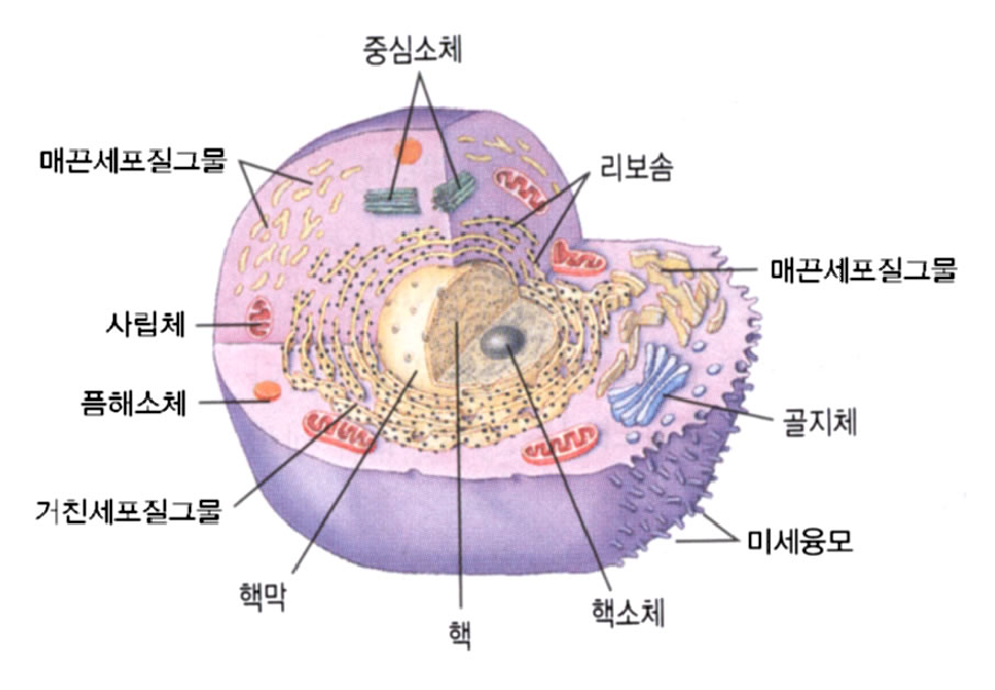 |
||
| 영문 | cell-mediated immunity | 한글 | 세포매개면역 |
|---|---|---|---|
| 설명 | 면역이란 신체를 외부의 물질로부터 보호하는 행위를 말한다. 여기에는 특이적 면역과 비특이적 면역의 두 가지가 있다. 비특이적 면역이라함은 특정한 물질에 관계하는 면역이 아니라 특정 대상이 없이 모든 외부 물체에 작용할 수 있는 면역을 말한다. 여기에는 소변의 흐름, 눈물의 흐름, 피부의 비투과성 등의 기계적인 것도 포함되고 피속에 돌아다니는 세포 중에서 비특이적으로 외부의 물질을 포식하는 세포들(예를 들면 큰포식세포(macrophage)의 활동도 포함이 된다. 세포매개면역이란 특이한 물질을 감지할 수 있는 세포를 생성하게 하여 그것으로 하여금 그 물질을 포식하게 하는 것을 말한다. |
||
| 영문 | nerve cell | 한글 | 신경세포 |
|---|---|---|---|
| 설명 | 신경세포는 올바른 신경전달을 위한 각 부분별로 나뉘어져 있다. 신경세포에서는 전해져오는 자극을 전기적인 신호로 바뀌어 보내거나 받게 된다. 이런 전기적인 현상은 각 신경세포내에 존재하는 각 이온채널(ion channel: ion이란 나트륨, 칼륨 등을 지칭하는 말들로써, 이들이 세포막에 의해 나뉘어질 때 생기는 전압차가 전기적 자극을 일으키고 유지하는데 결정적인 역할을 한다)들의 작용에 의해 이루어지게 된다. |
||
| 영문 | glia cell | 한글 | 아교세포 |
|---|---|---|---|
| 설명 | 신경세포 사이에서 그물구조를 이루며 이를 지지하는 조직. 신경아교세포는 신경모세포와 갈라진 아교모세포가 다시 여러 형태로 분화-성장한 것이다. 뇌실이나 척수중심관의 벽을 덮고 원주상 또는 입방형이며, 초기에는 유리면에 섬모가 있다. 대형세포는 별뇌실막세포는 아교세포라고 하며, 신경세포나 신경섬유 사이에 산재한다. 그 외에 희소돌기아교세포도 포함된다. |
||
| ACC | accommodation; acetyl coenzyme A carboxylase; acinic cell carcinoma; acute care center; adenoid cyst... |
|---|---|
| CEC | central echo complex; ciliated epithelial cell; Commission of the European Community |
| ECV | epithelial cell vacuolization; extracellular volume; extracorporeal volume |
| GEC | galactose elimination capacity; glomerular epithelial cell |
| HEP | hemolysis end point; hepatoerythropoietic porphyria; high egg passage [virus]; high-energy phosphate... |
| pigmented layer of ciliary body | The continuation of the pigment layer of the retina onto the posterior aspect of the ciliary body. Synonym: pigmented layer of ciliary body. (05 Mar 2000) |
|---|---|
| pigmented layer of iris | The double layer of pigmented epithelium on the posterior surface of the iris. Synonym: pigmented layer of iris. (05 Mar 2000) |
| pigmented layer of retina | The outer layer of the retina, consisting of pigmented epithelium. Synonym: ectoretina, stratum pigmenti bulbi, stratum pigmenti retinae, tapetum nigrum, tapetum oculi. (05 Mar 2000) |
| pigmented liver | A liver that contains pigment, such as occurs in Dubin-Johnson syndrome, haemochromatosis, long-standing malaria. (05 Mar 2000) |
| pigmented part of retina | pigmented part of retina |
| pigmented purpuric lichenoid dermatosis | An eruption comprised of lichenoid papules variously pigmented from the haemosiderin of the associated purpura; found on the legs, usually in men over 40 years of age. Synonym: Gougerot and Blum disease. (05 Mar 2000) |
| pigmented villonodular synovitis | <radiology> Monoarthritis, young adults, erosions on BOTH sides of joint (!), probably inflammatory, haemosiderin deposited in synovium, articular cartilage preserved (despite extensive marginal erosions; similar to gout), NO calcification Differential diagnosis: TB (associated atrophy of muscle and bone), rheumatoid arthritis (symmetrical), synovial sarcoma (with or without calcified; outside joint), synovial osteochondromatosis (12 Dec 1998) |
| congenital giant pigmented nevus | These large pigmented (often hairy) congenital nevi are important because of their increased risk (10 to 15%) of conversion into malignant melanoma. A biopsy can confirm if cells have turned malignant. Any change in a pre-existing nevus should prompt a physician evaluation. (27 Sep 1997) |
| nevus, pigmented | A nevus containing melanin. The term is usually restricted to nevocytic nevi (round or oval collections of melanin-containing nevus cells occurring at the dermoepidermal junction of the skin or in the dermis proper) or moles, but may be applied to other pigmented nevi. (12 Dec 1998) |
| synovitis, pigmented villonodular | Outgrowths of synovial membrane composed of villi and fibrous nodules characterised histologically by haemosiderin- and lipid-containing macrophages and multinucleated giant cells. It usually occurs in the knee. (12 Dec 1998) |
| calcifying epithelial odontogenic tumour | A benign epithelial odontogenic neoplasm derived from the stratum intermedium of the enamel organ; a painless, slowly growing, mixed radiolucent-radiopaque lesion characterised histologically by cords of polyhedral epithelial cells, deposits of amyloid, and spherical calcifications. Synonym: Pindborg tumour. (05 Mar 2000) |
| Malassez' epithelial rests | Epithelial remains of Hertwig's root sheath in the periodontal ligament. (05 Mar 2000) |
| microcystic epithelial dystrophy | Bilateral, symmetrical intraepithelial cysts in the central area of the cornea of healthy women, without hereditary predisposition. (05 Mar 2000) |
| polymorphic epithelial mucin | <protein> See episialin. (18 Nov 1997) |
| neoplasms, glandular and epithelial | Neoplasms composed of glandular tissue, an aggregation of epithelial cells that elaborate secretions, and of any type of epithelium itself. The concept does not refer to neoplasms located in the various glands or in epithelial tissue. (12 Dec 1998) |