| 영문 | rheumatic heart disease | 한글 | 류마티스심장병 |
|---|---|---|---|
| 설명 | 사슬알균감염 후 생기는 심장판막병이다. 원인은 A군 -용혈사슬알에 의한 인두염후 일종의 면역반응으로 발병한다. 진단은 존의 기준에 의한다. (1) 주요기준은 관절염 심장염(심장비대, 심장잡음, 심장기능상실 등) 무도증: 무당이 춤을 추는 것 같은 행동의 발작증세. 연변홍반: 빨간 테두리를 가진 피부병변은 피하결절(subcutaneous nodule): 피부 밑에 생긴 결절, (2)참고 기준은 열, 관절통, EKG상 PR연장: 심전도 소견 급성기 반응물질(예: ESR, CRP)의 상승, 류마티스열 치료는 페니실린으로 치료하고 심장의 후유증 또한 페니실린으로 예방한다. |
||
| 영문 | congenital heart disease | 한글 | 선천심장병 |
|---|---|---|---|
| 설명 | 선천적으로 심장의 구조에 이상이 있는 병. |
||
| 영문 | heart | 한글 | 심장 |
|---|---|---|---|
| 설명 | 심장은 가슴의 중앙에 위치하며 오른쪽과 왼쪽반으로 나눌 수 있다. 각각은 정맥에서 피가 들어오는 방과 동맥으로 피를 내보내는 실이 있다. 심장은 정중앙선에서 왼쪽에 놓여 있다. 심장의 2개의 심방과 2개의 심실, 그리고 심방사이의 심방중격, 심실사이의 심실중격이 존재한다. 심실은 혈액을 주로 순환시키는 힘을 내는 곳으로 강한 근육층이 존재를 한다. 반면에 심방은 심실에 들어가기전에 일시적으로 피가 모이는 곳으로 근육층의 발달이 미약하다. 심방과 심실사이에 삼첨판과 승모판, 심실의 유출로에 대동맥판과 폐동맥판이 있다. 이것들은 혈액이 흐르는 방향에 거슬러 진행하는 것을 막는 기능을 한다. 그리고 심장에는 심장의 근육에 혈액을 공급하는 관상동맥이 있다. 이 심장동맥은 심장을 쌓듯이 존재한다. 심장박동의 신호는 특수한 장치에 의해서 발생이 되고 특수한 전도계를 통해서 전체의 심장으로 전도된다. 심장박동의 신호는 우선 우심방에 위치하는 동방결절(SA node)라는 곳에서 우선 생긴다. 여기에서 보내는 전기적 신호가 바로 심장을 뛰게하는 신호인 것이다. 이 신호로 말미암아 심방이 우선 수축을 한다. 그리고는 심방과 심실사이에 위치하는 방실결절(AV node)라는 곳에 일정하게 정해진 통로없이 전체의 심방을 통해서 전달이 되고 이 신호를 받은 방실결절(AV node)는 히스 다발(His bundle)과 심장전도근육섬유를 통해서 심실의 심장근육에게 이 신호를 보내주어 심실근육을 수축하게 한다. 히스다발과 심장전도근육섬유는 전체의 심실의 심근섬유에 동시에 전기적 신호를 보내어 줄 수 있게 하는 일종의 전기줄과 같은 역할을 한다. 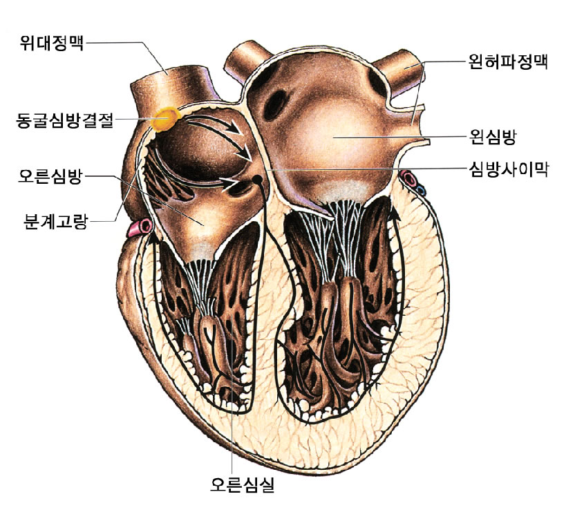 |
||
| 영문 | heart failure | 한글 | 심장기능상실 |
|---|---|---|---|
| 설명 | 몸의 조직이나 기관에서 대사에 필요한 만큼 충분한 양의 혈액을 공급할 수 없을 정도로 심장기능이 저하되어 있는 상태이다. 심장기능상실은 심근이 수축할 능력이 저하되었을 때나 심장에 심박출을 하기 위한 압력이 정상보다 증가되어 정상의 심장의 수축으로는 충분한 양의 혈액을 공급할 수 없는 경우, 그리고 심장근육, 심장에 걸리는 압력은 정상이나 심장박동의 이상에 의해서 정상적인 수축이 불가능할 경우에 생긴다. |
||
| 영문 | pacemaker(of heart) | 한글 | 심장박동기 |
|---|---|---|---|
| 설명 | 심장의 전기적 자극이 병적인 상태로 발생하지 않거나, 혹은 심실로 잘 전해지지 않을 때 사용한다. 일시적 심장박동기와 영구적 심장박동기가 있으며, 각기 쓰이는 용도는 병에 따라 다르다. 요즘에 나오는 심장 박동기는 건전지의 수명도 반영구적이며, 밖에서 조정할 수 있고, 운동이나 스트레스 상황에 대한 심장의 빠른 운동에도 잘 적응할 수 있도록 만들어져 있다. |
||
| HI | half-scan with interpolation; head injury; health insurance; hearing impaired; heart infusion; hemag... |
|---|---|
| IHD | Ischemic Heart Disease = Coronary Heart(Artery) Disease = Atheroscler... |
| JVP | [POMD P 49 - 52] 1) Jugular Vein Pressure 2) Jugular Venous Pulse ... |
| AHD | acquired hepatocerebral degeneration; acute heart disease; antihyaluronidase; antihypertensive drug;... |
| CHD | Chediak-Higashi disease; childhood disease; chronic hemodialysis; congenital or congestive heart dis... |
pulmonary pleura
| MUGA scan | This noninvasive test uses radioactive tracers to delineate the hearts chambers and major vessels. It may be used to detect a heart attack, heart muscle function and coronary artery disease. The patient receives a radioactive tracer by injection (into a vein) and then the heart is imaged using a gamma camera. The heart is imaged before and after exercise. This test may be used to detect and evaluate atrial septal defect, dilated cardiomyopathy, congestive heart failure, cardiomyopathy, Lyme disease (secondary), mitral stenosis and superior vena cava syndrome. (27 Sep 1997) |
|---|---|
| CT scan | <investigation, procedure, radiology> A special radiographic technique that uses a computer to assimilate multiple X-ray images into a 2 dimentional cross-sectional image. This can reveal many soft tissue structures not shown by conventional radiography. Scans may also be dynamic in which a movement of a dye is tracked. Cuts may be 5 or 10 mm apart or, in some instances even further apart. A special dye material may be injected into the patients vein prior to the scan to help differentiate abnormal tissue and vasculature. The machine rotates 180 |
| scan | A type of imaging, for example ultrasound, MR, CT, scintigram. (16 Dec 1997) |
| scan rate | <microscopy> The number of horizontal-scan lines per frame and vertical scans per second that are repeated in video, for example, 525/60, 625/50. In 525/60, 2: 1 interlaced video, the V scan is repeated at the field rate (which is half of the frame rate for 2: 1 interlaced video) so that 525 H scans take place 30 times a second. The H-scan rate is therefore 525 x 30 = 15.75 kHz. With 525/60, 1: 1 interlace, the H-scan rate would be twice this value. (05 Aug 1998) |
| sector scan | In ultrasonography, a system in which the transducer or transmitted ultrasound beam is rotated through an angle, resulting in a pie-shaped image. (05 Mar 2000) |
| slow scan | <microscopy> A system of video scanning in which the time used to read each line has been increased in comparison to standard video. The bandwidth needed to faithfully transmit or record the signal is reduced in inverse ratio to the scanning time. Slow scan allows the video signal to be transmitted over a telephone line, or line scans to be registered on a chart recorder. (19 Jan 1998) |
| nuclear bone scan | A nuclear medicine test that involves the introduction of a radioactive compound into the blood stream. The radioactive compound acts as a tracer and allows for the imaging of the bony skeleton. (27 Sep 1997) |
| nuclear scan: adrenals | A nuclear scan that images the adrenal glands after a radioactive tracer is injected into the bloodstream. This test is useful in detecting a pheochromocytoma, particularly if it not within the adrenal gland. (27 Sep 1997) |
| duplex Doppler scan | A method of visualizing and selectively assessing the flow patterns of peripheral arteries and veins using ultrasound imaging and pulsed Doppler. (05 Mar 2000) |
| testicular scan | <radiology> Tc-99m pertechnetate 30 mCi, interpretation: torsion = cold defect, epididymo-orchitis = hot spot, trauma = hot or cold (12 Dec 1998) |
| thyroid scan | A picture taken of the thyroid gland after radioactive iodine is taken by mouth. (12 Dec 1998) |
| EMI scan | Historically, the name commonly used for computed tomography of the head, the technique devised by Hounsfield, who was a scientist at EMI, an English electronics firm. (05 Mar 2000) |
| liver scan | <investigation> A way of visualising the liver by injecting into the bloodstream a trace dose of a radioactive substance which helps visualize the organ during X-ray. (09 Oct 1997) |
| liver-spleen scan | <radiology> Tc-99m sulfur colloid or albumin colloid, particles less than 1 m, dose = 4-8 mCi Distribution, liver 85%, spleen 10%, bone marrow 5% Findings, liver: hot / cold, spleen: hot / cold (12 Dec 1998) |
| abnormal heart chamber dimensions | <radiology> Left ventricular volume overload, left ventricular hypertrophy, right ventricular volume overload, right ventricular hypertrophy, fixed subvalvular aortic stenosis, hypoplastic left/right ventricle; common ventricle, congestive cardiomyopathy (12 Dec 1998) |